

|Videos|December 22, 2017
Classification Guidelines for Gastric/GEJ Cancers
Advertisement
Transcript:
Yelena Y. Janjigian, MD: In esophageal cancer, there are 2 distinct subtypes. The esophagus cancer, esophageal adenocarcinoma, and esophageal squamous cell cancer. Historically, we’ve known that these tumors are very distinct and they have distinct risk factors and biologic behavior. But unfortunately, historically, these patients also used to be put together in similar clinical trials. As the field has evolved, we know that squamous cell of the esophagus is molecularly quite distinct from adenocarcinoma of the esophagus. And as patients become more aware of health risk factors of smoking or alcohol intake, the overall incidence of squamous cell cancers is declining in the Western world. And so, esophageal squamous cell cancer is quite rare now in the United States and the West.
In Asia, esophageal squamous cell cancer is quite common still, particularly in parts of China where it’s related to HPV, or human papilloma virus. Certain dietary exposures, such as pickled foods, smoking, and drinking alcohol, are the major risk factors. As I mentioned, in the United States it’s exceedingly rare. Now, squamous cell of the esophagus is mostly related to smoking, alcohol intake, or prior radiation therapy in patients who are survivors from lymphoma or breast cancer.
Adenocarcinoma of the esophagus is biologically very similar to the GE-junction tumors. And so, within the subtype, these are tumors that are targeted together, at least in metastatic trials for locally advanced resectable tumors. Their approaches for the operation are slightly different. But again, the distinction is really between squamous disease and the adenocarcinomas. Within adenocarcinomas, esophageal and GE junction tumors, and even approximal stomach tumors, all behave similarly.
Histologically within adenocarcinomas, there are different ways these appear under the microscope, and these are the WHO classifications and the Lauren classification. What we know about adenocarcinomas is that there are different types of cell that resemble a gland, or glandular cell, from where the cancer started at the mucinous part of the stomach and a GE junction. The closer the cell still resembles the architecture of a normal gland, the more well or moderately differentiated it is. And so, within the WHO classification, there are different grades and differentiation; moderately to poorly differentiated cells; and beyond that, poorly differentiated cells with signet ring cell histology.
These are more historic classifications within pathology. Within metastatic tumors, it may give us a sense of the tumor behavior and their metastatic pattern, but it doesn’t really change systemic chemotherapy per se. It’s just a different way that we used to classify and think about these tumors. Now the field is moving more beyond that, beyond anatomy, beyond just a crude appearance of the cell under the microscope on an H&E (hematoxylin and eosin) slide, and into the molecular subtyping, which also falls into this classification, the Lauren classification, nicely.
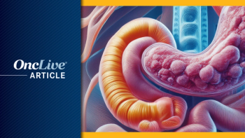
Within GE junction tumors, we further subdivide them. The length of the esophagus in an average human being is approximately 42 cm, and beyond that is where the GE junction and stomach starts. And so, in the United States actually, the most common subtype of adenocarcinoma of the esophageal gastric region are the GE junction tumors.
For the GE junction tumors specifically, we are further subdividing them into 3 different subtypes by the Siewert classification. Dr. Siewert is a surgeon who developed this classification, and it helps us to determine what type of operation the patient needs. It’s mostly important for tumors that have not metastasized, as the Siewert classification helps us determine where in the GE-junction the tumor is located. For Siewert I, those are tumors that are closest to the esophagus, and Siewert III are the tumors that are located at the very bottom of the GE-junction extending into the stomach. Anything in between is Siewert II, and for Siewert I and II tumors we tend to approach them as esophageal cancer.
For preoperative treatment chemoradiation, it is important to help clear the margins and get rid of the disease completely at the time of surgery. For Siewert III tumors, although chemoradiation can also be applied, historically we’ve treated them as proximal stomach cancer. And so, the type of operation they usually need is a total gastrectomy, and they receive preoperative chemotherapy alone.
Transcript Edited for Clarity
Advertisement
Related Content
Advertisement


Latest CME


Advertisement
Advertisement
Trending on OncLive
1
Single-Center, Retrospective Data Show Low Rate of Lifileucel Infusion Following Referral in Advanced Melanoma
2
Long-Term Cilta-Cel Data Show Low Rates of PFS Events in Standard-Risk R/R Myeloma
3
Real-World Data Support Clinical Benefit With Lifileucel in Previously Treated Advanced Melanoma
4
Dr Riedell on the Long-Term Efficacy of Tisa-Cel in R/R Follicular Lymphoma
5