
|Articles|December 12, 2018
- December 2018
- Volume 7
- Issue 6
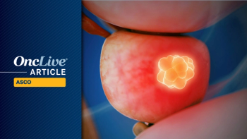
Focal Therapy Evolves Into More Precise Treatment in Prostate Cancer
Historically, the management of localized prostate cancer has centered on removal or ablation of the entire gland with either extirpative surgery or radiation. However, the subsequent unacceptable adverse events profile can be devastating and life altering for many patients.
Advertisement
Brian J. Miles, MD
For more than a generation, the notion of lumpectomy instead of radical mastectomy for breast cancer was met with scorn and treated as heresy. Now, it is the standard of care and has saved the physical QoL for many women. Prostate cancer (PCa), which is similar to breast cancer in many ways, has also been considered not amenable to focal therapy as a form of lumpectomy. This is because of the size of the prostate, its relative inaccessibility, the fact that PCa is often multifocal, and concerns that focal therapy may rule out some treatment options if the cancer recurs. However, with the recent FDA clearance of high-intensity focused ultrasound (HIFU) in prostate disease, this therapeutic option has become viable. HIFU has been used for decades in Europe with excellent oncologic control and acceptable AEs.
The potential for prostate lumpectomies expanded notably in June 2018, which is when the FDA cleared the Focal One HIFU device as the first medical apparatus designed specifically for focal treatment of the prostate, although earlier devices made this procedure possible. As with all technological advances, long-term studies and clinical data will ultimately determine whether Focal One distinguishes itself as a uniquely useful instrument. Nevertheless, its arrival is significant due to the degree of precision with which diseased prostate tissue can be ablated while sparing healthy tissue and thus reducing AEs.
The Focal One device provides precision capability to treat the prostate by integrating magnetic resonance and 3D biopsy data with real-time ultrasound imaging—a process known as fusion. This melding of advanced imagery and patient diagnostic data provides a detailed 3D view of the prostate on a large monitor in real time, enabling direct highintensity ultrasound waves to ablate the targeted area within the prostate.
Because this fusion of technologies allows for improved targeting of diseased tissue during treatment planning, the margins around the contours of that tissue can be drawn more concisely than the margin contours required by less-precise procedures used in the past, thus eliminating the need to remove larger volumes of prostate tissue. In addition, this advancement in focal ultrasound prostate tissue ablation enables clinicians to enact a protocol that respects the maxim “right patient, right treatment, right time,” because the focal HIFU therapy can be customized for each patient and for each clinical scenario.
Figure 1. German Study: Survival Rates for Patients With Localized PCa Treated With HIFU1
Long-Term Outcomes
During the procedure the device’s proximity to the rectal wall is constantly monitored for patient safety. The robotic HIFU device automatically readjusts itself and, if necessary, stops transmission of the high-frequency waves anytime they are too close to the rectal wall. This aids in avoiding rectal injury.Future long-term studies are hoped to provide data needed to fully analyze the potential of focal HIFU therapy as an alternative to radiation and radical prostatectomy for treatment of localized PCa in the United States. European researchers have published positive peer-reviewed studies of the use of this technology for whole gland ablation.
Here is a brief review of 2 such studies: In 2013, the Journal of Urology published a German study of 704 patients treated with HIFU, in which co-investigators Stefan Thüroff and Christian Chaussy concluded that long-term follow-up with HIFU therapy demonstrated a high overall rate of cancerspecific survival (99%) and 10-year salvage treatment-free rates of 98% in low-risk, 72% in intermediate-risk, and 68% in high-risk patients. “Advances in HIFU technology and clinical practice, as well as the use of neoadjuvant transurethral prostate resection, allow the complete treatment of any size prostate without inducing metastasis,” the authors wrote.1
The study assessed patients with primary localized PCa (T1-2, N0, M0, and prostate specific antigen [PSA] at first diagnosis <50 ng/mL) and follow-up longer than 15 months. Patients with previous long-term androgen deprivation therapy (ADT), locally advanced PCa, or any therapy influencing PSA were excluded from the study.
Among the study participants, 78.5% had intermediate- or high-risk disease. The mean follow-up was 5.3 years (range, 1.3-14). Metastasis-free survival was 95%. PSA nadir and Gleason score predicted biochemical failure, and AEs were moderate. The OS of the patient population was identical to current local Bavarian population survival statistics (Figure 1).1 Of the total patients, 38.4% were intermediate risk and 40% were high risk based on stratification tools of the 1990s and early 2000s. The study also concluded that the rate of low-risk patients being able to avoid salvage therapy was exceptionally high, based on long-term follow-up.
A second European study investigated HIFU-based whole-gland ablation of localized PCa in 1002 patients. This was led by Sebastien Crouzet in France, with results published by the European Association of Urology. Participants were patients treated from 1997 to 2009.2 The 10-year PCa-specific survival and metastasis-free survival rates were 97% and 94%, respectively.
Also, severe incontinence and bladder outlet obstruction decreased with refinement in the technology, from 6.4% and 34.9% to 3.1% and 5.9%, respectively. Limitations included that the study was a single-arm study without a comparison group. Researchers concluded that HIFU is a “potentially effective treatment” of localized PCa, with a low PCa-specific mortality rate and a high metastasis-free survival rate at 10 years, as well as acceptable morbidity. The 10-year OS rate and PCa-specific survival rate were 80% and 97%, respectively (Figure 2).2 About two-thirds of the patients were determined to be intermediate or high risk during an era of frequent understaging.
Advancing Treatment Options
In addition to this European research, the University of Miami Miller School of Medicine conducted a study of functional and oncologic outcomes resulting from HIFU for focal treatment of PCa. Preliminary results were presented at the Southeastern Section of the AUA conference in March 2018. Researchers at the University of Miami also introduced the FoR-UsA Registry (Focal Robotic Ultrasound Ablation Registry) in February, creating the first registry in the United States to specifically track baseline and long-term follow-up data on patients who have undergone focal therapy for prostate tissue ablation.In Europe and in countries elsewhere, HIFU has been used to treat more than 50,000 patients. The accumulating data have established focal HIFU as a safe and effective therapy with statistically significant survival rates in the 3-to-5-year window. Further, the data from the 2 European studies mentioned here document positive outcomes at the 5-to-10-year follow-up points.1-3
As these studies and other research show, focal HIFU is taking its place as a viable option among focal therapies for patients with localized PCa. Continued international research, US studies, and a US HIFU registry data collection are poised to bolster the case for making patients aware of this option.
Focal Laser Ablation
Focal therapies are aimed at providing a middle ground for patients with localized PCa who wish to avoid the potential life-altering AEs that come with radical prostatectomy or radiation on the one hand or the stress of active surveillance on the other. Besides HIFU, laser ablation and cryotherapy also are a part of the tool set for achieving this goal.Focal laser ablation (FLA) uses commercial laser fibers, generally 980-nm diode lasers. This method has been studied and used since it was determined to be a feasible alternate treatment approach by Lindner et al in 2009.4 FLA uses MRI guidance to treat regions of interest (ROIs) that are biopsy-confirmed areas of cancer. Thermal ablation of the cancer is then carried out, often under MRI thermometry control.
In the 2009 study, 12 men were treated with transrectal ultrasound fused with biopsy-proven MRI-determined ROIs. Target ablation was monitored with thermal sensors and contrast-enhanced ultrasound, and AEs, including the impact on voiding and erectile function, were evaluated in the aftermath.
The median MRI cancer volume was 0.25 cc (range, 0.1-0.99). Temperature probes were placed at the ablation zone boundary to ensure temperatures of ≥55oC were attained at the borders. Follow-up MRIs at 7 days revealed the median treatment lesion volume was 2.2 cc (range, 0.3-4.0), creating an ablated lesion that was an average of 12.25 times the tumor size. The overall median preand posttreatment MRI overlap was 53%, although better (83%) in the last 4 patients, demonstrating consistent imaging.
In the posttreatment follow-up, 10 core biopsies with 2 additional cores in the treatment zone revealed half of the patients to have completely negative biopsies; 2 of these patients had negative biopsies in the treatment zone but a positive low-volume core in the contralateral side. Four patients were treatment failures, with 3 having poor MRI overlap.
The authors concluded that image-guided focal photothermal ablation of low-risk and low-volume PCa is feasible, with minimal AEs; however, they said further studies would be required to demonstrate the efficacy of this treatment.
In a follow-up study, Lindner et al reported in 2010 on 4 men treated with FLA 7 days prior to radical prostatectomy.5 The study was designed to understand how well MRI represents true treatment effect and how effective FLA was in this group. The authors found that on “vital stain” pathologic evaluation, MRI-ablated volumes were 1.1 times greater than the pathology findings. They felt they had demonstrated that it was possible to fully ablate a specified volume in preoperatively chosen zones, leaving no viable cells. No mention was made of cancer cells at the borders of the treatment zone. Nonetheless, it does suggest the need for a safety zone around MRI-defined ROIs.
In 2015, Lepor et al reported on 25 men treated “in bore” (ie, in the MRI chamber) with FLA. They used a 600-μm laserdiffusing fiber with a 15-mm tip passed through a cooled catheter system, meant to keep fiber from overheating.6 These 25 patients had 28 ROIs treated, with 26 showing “no evidence of PCa” in the treatment zone.
Another in-bore study was reported in 2016 by Eggener and colleagues.7 In this phase II evaluation of MRI-guided FLA, at 3 months post treatment, 96% of the total 27 men had no cancer in the treatment zones on image-guided biopsies. A 15-watt fiber with reduced power was employed, and the treatment duration was 60 to 120 seconds at 6- to 15-watt bases on the intended size of the treatment zone. The target ablation temperature achieved was reported as a minimum of 60o C. The mean procedure duration was 197 minutes (range, 138-302), and the mean ablation time 320 seconds. The mean duration of treatment for the final 10 patients was 160 minutes. In 18 men, 1 MRI lesion with cancer confirmed by a biopsy was targeted, and 2 lesions were targeted in 9 patients. Follow-up biopsies at 1 year revealed cancer in 37% (10 men), including 11% (3 men) with cancer in the treatment zone.
In an effort to make FLA a bit more user friendly, Natarajan and Marks reported, in 2017, on 10 patients treated in the clinic under local anesthesia using MRI/ transrectal ultrasound fusion technology.8 For safety purposes, a temperature probe was near the rectal wall and in the ablation zone. The treatment involved a 15-watt laser fiber activated at 13.75 watts for a treatment time of 3 minutes (less if the rectal probe reached 42o C or the parallel probe reached 70o C). The patients tolerated the procedure well, with a maximum pain score of 5 reported during treatment. The mean treatment zone volume was 4.3 cc (range, 2.1-6.0) and the procedure took an average of 95 minutes (range, 71-105) to accomplish. At 6 months after treatment, fusion biopsies revealed only 3 patients were cancer free in the treatment zone. Of those deemed failures, 4 had Gleason score 7 disease present in the ablation area with maximum core length of 3 to 9 mm.
In these studies, QoL was generally well preserved. International Index of Erectile Function scores were unchanged or improved, and sexual function was unchanged or returned to baseline within 6 months. Most men were catheter-free at discharge post procedure, and all patients were catheter-free within 3 days post procedure. Issues such as potential hematuria, perineal ecchymosis, etc, were minimal.
Figure 2. French Study: Survival Rates for Patients With Localized Pca Treated With HIFU2
Focal Cryoablation
All of these published studies of FLA support the concept of focal ablation for MRI-confirmed PCa. Nonetheless, they also reveal that oncologic control within the ablation zone can be haphazard, that issues with MRI definition of adequate treatment remain,9 and that treatment times can vary greatly, suggesting a steep learning curve. As MRI technology and localization of significant cancer improve and carefully conducted trials are carried out, the potential of this FLA therapy will become better defined.The goal for focal or hemiablation cryosurgery in properly selected men is to reduce adverse consequences from more traditional therapy, such as urinary incontinence, stricture disease, bowl toxicity, and erectile dysfunction, while providing comparable cancer outcomes. All patients undergo a double freeze—thaw cycle during treatment with active thawing. Typically, 3 treatment probes are used along with 2 to 3 temperature probes to monitor treatment areas and the external sphincter. A urethral warming catheter is always utilized, and all focal patients typically are discharged on the same day. A suprapubic tube is not utilized, and most patients go home with a standard Foley catheter for 3 to 5 days. Patients have minimal discomfort and use of narcotics is rare. Monitoring is similar to that provided for radiation therapy, with PSAs every 3 months for a year and then every 6 months until the physician determines it is appropriate to stop. Repeat biopsies are not performed unless biochemical recurrence (BCR) is suspected based on the ASTRO definition (detectable or rising PSA value after surgery that is ≥0.2 ng/ml with a second confirmatory level ≥0.2 ng/ml).10
Focal therapy, in general, started with cryotherapy/ cryosurgery. In 2007, Onik et al reported on the “male lumpectomy” using focal cryotherapy.11 Their protocol started in 2000 and reported on 93 men, 55 of whom had not suffered radio recurrent disease, with greater than 1 year of follow-up treated with focal cryoablation. Of these 55, 95% had a stable PSA. Twenty-nine had a biopsy at 1 year, and 26 were negative. The 3 remaining patients had a positive biopsy, and these were in untreated areas of the gland. Potency was preserved in 85%, and only 1 patient reported urinary incontinence, 2 pads daily. A year later, the investigators updated their reporting on 48 of the men; this group now had at least 2 years of follow-up. No local recurrences were found in the treated areas, nor was cancer found in the gland, and all biopsy findings were negative among the 26 patients with a stable PSA level who had undergone routine biopsy at 1 year. Ninety-four percent of the men had stable PSAs, with potency still in the 85% range, and none experienced incontinence.
One of the earliest reports on focal cryotherapy was from Bahn et al in 2006.12 In this study, 31 men with a mean age of 63 years underwent focal or hemiablation therapy. Follow-up for these patients was PSA monitoring every 3 to 6 months. Most significantly, patients underwent repeat prostate biopsies at 6 months, 12 months, 24 months, and 5 years. At a mean follow-up of 70 months, and according to the ASTRO definition, 92.8% of patients were still biochemically disease free and 96% had a negative biopsy rate. A potency rate of 88.9% was achieved: 48.1% of men not requiring any medical assistance for erections and 40.7% requiring oral pharmaceutical therapy for erections. There were no reports of stress urinary incontinence.
A follow-up study by Bahn in 2012 with 73 men showed encouraging outcomes as well.13 Complete follow-up was obtained in 70 patients, and all patients had repeat biopsies at 6 and 12 months and then yearly. Seventyfive percent of the patients had negative biopsies, and complete continence and potency was achieved in 100% and 86% of patients respectively.
Ward and Jones reported in 2011 on a large series of men from the COLD Registry, a national cryo online database with voluntary reporting from many urologists throughout the country.14 They found 1160 men managed with focal cryoablation. Most men (74%) had a Gleason score of ≤6, and 87% had stage ≤cT2b. Focal therapy increased every year from 1999 to 2007.14 BCR at 2 years was approximately 24%, similar to the whole gland results. Biopsy was only performed for men with a rising PSA. Of the group, 163 (14.1%) were biopsied and 43 of these (26.3%) were positive. No data on whether the cancer was in the treated zone were provided. Morbidity was low, consistent with the other studies, although new-onset erectile dysfunction occurred in 42%. A big issue with this database is reliance on individual urologists providing all the necessary treatment and follow-up data, which, in general, has been haphazard in this registry.
Conclusions
Another focal study from Barqawi et al from 2014 demonstrated a negative biopsy in 81% of patients.15 Sixty-two patients were treated, and no patients experienced postoperative erectile dysfunction or stress urinary incontinence. These patients were diagnosed with 3D mapping biopsy using triplane transrectal ultrasonography.Although all of these studies on early focal therapies— HIFU, cryo, and laser—are optimistic and generally enthusiastic, good long-term studies would be welcome. Unfortunately, in many respects, the “horse has already left the barn.” Patients want HIFU treatment, and practicing urologists are increasingly embracing it. As stated by Ward and Jones, “Although the prognosis for PCa is now very different from what it was 20 years ago, the aggressiveness of our treatment has not changed commensurately,” and furthermore “the concept of organ-sparing therapy went from improbable to plausible, and the explosion of its use…shows its appeal to the urologic community.”14
A randomized multicenter control trial between these 3 modalities would be optimal, but it is most likely not feasible. Certainly, single-center studies of each option would be valuable and ongoing multicenter trials would be helpful. However, we believe a well-designed registry trial with defined pre— and post–focal therapy data that must be collected will be helpful. Failure will have to be well defined and dependence on the ASTRO definition of a BCR used in patients who have undergone radiation treatment will undoubtedly need to be replaced by either PSA kinetics, a single rise of greater than a “certain” percent, possibly PSA density, genomic profiling, or a combination of those. A biopsy requirement at 1 year, etc, will generally not be supported by urologists or patients unless required by laboratory or clinical data. We need to adapt to our current reality and move forward with the knowledge of where we are and what we can do. Focal therapy could become the male lumpectomy for appropriate men.
References
- Thüroff S, Chaussy C. Evolution and outcomes of 3 MHz high intensity focused ultrasound therapy for localized prostate cancer during 15 years. J Urol. 2013;190(2):702- 710. doi: 10.1016/j.juro.2013.02.010.
- Crouzet S, Chapelon JY, Rouvière O, et al. Whole-gland ablation of localized prostate cancer with high-intensity focused ultrasound: oncologic outcomes and morbidity in 1002 patients. Eur Urol. 2014;65(5):907-914. doi: 10.1016/j.eururo.2013.04.039.
- Prostate cancer: focused ultrasound treatment for the prostate is available in the United States. Focused Ultrasound Foundation. Published May 9, 2018. Accessed November 11, 2018. fusfoundation.org/diseases-and-conditions/oncological/prostate-cancer
- Lindner U, Weersink RA, Haider MA, et al. Image guided photothermal focal therapy for localized prostate cancer: phase I trial. J Urol. 2009;182(4):1371-1377. doi: 10.1016/j.juro.2009.06.035.
- Lindner U, Lawrentschuk N, Weersink RA, et al. Focal laser ablation for prostate cancer followed by radical prostatectomy: validation of focal therapy and imaging accuracy. Eur Urol. 2010;57(6):1111-1114. doi: 10.1016/j.eururo.2010.03.008.
- Lepor H, Llukani E, Sperling D, Fütterer JJ. Complications, recovery, and early functional outcomes and oncologic control following in-bore focal laser ablation of prostate cancer. Eur Urol. 2015;68(6):924-926. doi: 10.1016/j.eururo.2015.04.029.
- Eggener SE, Yousuf A, Watson S, Wang S, Oto A. Phase II evaluation of magnetic resonance imaging guided focal laser ablation of prostate cancer. J Urol. 2016;196(6):1670- 1675. doi: 10.1016/j.juro.2016.07.074.
- Natarajan S, Jones TA, Priester AM, et al. Focal laser ablation of prostate cancer: feasibility of magnetic resonance imaging-ultrasound fusion for guidance. J Urol. 2017;198(4):839- 847. doi: 10.1016/j.juro.2017.04.017.
- Eggener SE, Yousuf A, Watson S, Wang S, Oto A. phase ii evaluation of magnetic resonance imaging guided focal laser ablation of prostate cancer. J Urol. 2016;196(6):1670- 1675. doi: 10.1016/j.juro.2016.07.074.
- Thompson IM, Valicenti RK, Albertsen P, et al. Adjuvant and salvage radiotherapy after prostatectomy: AUA/ASTRO Guideline. J Urol. 2013;190(2):441-449. doi: 10.1016/j.juro.2013.05.032.
- Onik G, Vaughan D, Lotenfoe R, Dineen M, Brady J. “Male lumpectomy”: focal therapy for prostate cancer using cryoablation. Urology. 2007;70(suppl 6):16-21. doi: 10.1016/j.urology.2007.06.001.
- Bahn D, Silverman P, Lee F Sr, Badalament R, Bahn ED, Rewcastle JC. Focal prostate cryoablation: initial results show cancer control and potency preservation. J Endourol. 2006;20(9):688-692 doi: 10.1089/end.2006.20.688.
- Bahn D, de Castro Abreu AL, Gill IS, et al. Focal cryotherapy for clinically unilateral, lowintermediate risk prostate cancer in 73 men with a median follow-up of 3.7 years. Eur Urol. 2012;62(1):55-63. doi: 10.1016/j.eururo.2012.03.006.
- Ward JF, Jones JS. Focal cryotherapy for localized prostate cancer: a report from the national Cryo On-Line Database (COLD) Registry. BJU Int. 2011;109(11):1648-54. doi: 10.1111/j.1464-410X.2011.10578.x.
- Barqawi AB, Stoimenova D, Krughoff K, et al. Targeted focal therapy for the management of organ confined prostate cancer. J Urol. 2014;192(3):749-753. doi: 10.1016/j. juro.2014.03.033.
Articles in this issue
over 7 years ago
Immunotherapy Changes Early-Stage Urothelial Carcinoma Paradigmover 7 years ago
San Diego Urology Group Enjoys Growth in a Tough MarketAdvertisement
Related Content
Advertisement


Advertisement
Advertisement
Trending on OncLive
1
ASCO 2026 Ushers In a New Era for Pancreatic Cancer, Expands Targeted Therapy Options Across the GI Spectrum
2
Targeted Therapies, ADCs, and Oral SERD Combos Expand Treatment Possibilities Across Breast Cancer Settings
3
Previewing Key Myeloma Presentations to Watch at EHA 2026: With Joshua Richter, MD
4
Generic Ga-68 PSMA-11 Radiodiagnostic Nets FDA Approval in Prostate Cancer
5