Johannes Schetelig, MD, MSc, discusses phase 3 data for post-transplant cyclophosphamide vs ATLG as GVHD prophylaxis in hematologic malignancies.
Nichon L. Grupka, MD, discusses advances in hematopathology, specifically mutations as prognostic indicators, disease-associated versus disease-causing mutations, and advice for physicians who are working to navigate the field.
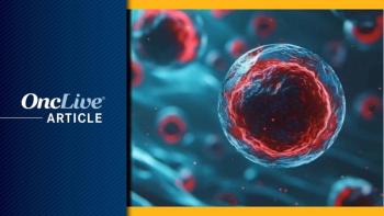
The 2016 published revision of the World Health Organization’s (WHO) classification of tumors and hematopoietic and lymphoid tissues provided pathologists with refinements to diagnosis, prognosis, and therapeutic approaches for myeloid neoplasms, but there is still much to discern, according to Nichon L. Grupka, MD.
“Now, there are prognostic implications for therapy and potentially classification. Though a lot of the criteria are the same, all of the myelodysplastic syndrome (MDS) categories have been altered to streamline the process,” said Grupka. “That includes more specific wording and the incorporation of new technology.”
Pathologists are now working to define what role these technologies, such as next-generation sequencing, will have in the classification and identification of disease states. She adds that pathologists are still working to understand the relationship between a number of mutations and malignancies.
In an interview during the 2018 OncLive® State of the Science SummitTM on Hematologic Malignancies, Grupka, a pathologist at Novant Health Thomasville Medical Center, discussed advances in hematopathology, specifically mutations as prognostic indicators, disease-associated versus disease-causing mutations, and advice for physicians who are working to navigate the field.Grupka: The biggest advancement is the release of the 2016 WHO classification; that incorporates a whole area of somatic mutations. We’d like to use next-generation sequencing to determine if a patient who has an abnormality in their genome has MDS. We’re still figuring out if we can use this technology to define that criteria.
Subsequently, what do we do with that information? That can be confusing for a lot of people because older people who do not have MDS, or any hematologic malignancy, can also possess these mutations. Some of those patients may even be cytopenic. The challenge is taking all of this technology and information and incorporating it into a body of knowledge that is constantly changing. We know the basic entities and, because of the WHO, a few additional criteria have been recognized. It's difficult if you've been practicing for a long period of time.
Everyone is busy seeing patients, so finding the time to sit down and read the revised guidelines can be difficult. That's where I see my role as a pathologist coming in. If a patient has a molecular mutation, we can use these new technologies to classify the patient. Sometimes, I can outright call it MDS, and sometimes I can't. Oftentimes, it’s a phone call to relay the information and describe what's missing from the criteria that's required for MDS.
A lot of these mutations in MDS are not necessarily specific to a particular disease entity. For instance, if someone has a t(9;22), a Philadelphia chromosome, they are likely to have chronic myeloid leukemia. However, if they have an SF3B1 mutation, the patient could have chronic lymphocytic leukemia (CLL) or MDS. The same is true with the BRAF mutation. We can see that in nonhematologic malignancies and hairy cell leukemia.
As I said, some mutations are not specific to a malignancy, and the presence of one may not mean anything for your patient at that time. It might mean that they have a predisposition to a malignancy downstream, or it might mean they have a predisposition to something nonhematopoietic, like a propensity for arteriosclerosis. It’s a vast field, and it's changing rapidly. Plasma cells will likely play a role in the field, as well. Patients with an SF3B1 mutation typically are associated with a very specific morphologic pattern. Patients with MDS who are associated with a ring sideroblast, oftentimes have a [different] disease course. It really depends on the context of that particular disease as opposed to it showing up in a patient with CLL. Some people have a TET2 mutation, and a lot of these patients have a neutral prognosis. Is that actually a mutation that allows for other mutations to accumulate? Does its presence predispose to other conditions, or does it potentiate other conditions? That's the difficulty because most of them are not necessarily very specific.Our genes have uniqueness, and we can have what's called polymorphisms. There’s the standard sequence of base pairs and DNA. Some people can have diversions, but it’s not necessarily disease-causing mutations. Usually you can sample bone marrow so you can determine whether or not it’s constitutional. You can take a buccal swab and sequence the DNA to see if they possess that polymorphism in an area of the body that isn’t suspected to have disease. Then you can determine whether or not it’s just a part of their makeup.
There’s always potential for this, so it should be kept in mind as a possibility. There are people who carry these mutations as family predispositions, so they were born with it. It’s not because they were exposed to something, or a cell mutated wrongly; it’s simply their germline DNA. Oftentimes, you see these GATA2 deficiencies and RUNX1 mutations in people who had unexplained thrombocytopenia for years; it’s not necessarily that they're in a disease state.
Now interestingly, it does predispose them to develop leukemia. They're at least 35% more likely to develop AML in their lifetime as opposed to an age-related cohort of patients who do not possess that mutation. Then again, they have lived with this cytopenia for a large portion of their lives. Many of these people don't get diagnosed until their 40s and 50s. That goes for one or both of their parents, and potentially another family member, as well.
While these mutations are disease-associated, they're not necessarily disease-causing; it really depends. It may be what we call a second head if you have a RUNX1 mutation and then something else comes along to activate it. A very large gene such as ASXL1 may contain a mutation; however, if it is small enough—a missense mutation—it may not express itself. It does potentially predispose people. As we get older, we naturally gain mutations; it’s a natural part of development. Ten percent of people in their 70s could have mutations in their genes that aren’t in a disease state.
If someone who had a mutation earlier gains more mutations as they get older, that person could potentially develop something. The people who have mutations have a less than 1% chance of developing a hematologic malignancy compared with the normal population. It's complicated and its difficult, but these are good data to know.
We know that if we have a patient with this type of mutation, in this gene at this allele frequency, we can have this expectation. If we test again and we find this specific mutation, we can determine the significance of it. You have to balance what's practical in its application and what you can readily test for. Although the technology is great, it’s expensive. That may come down in time. The easiest thing to do is look at a table because it breaks it down, so you’ll be able to look at the categories. If you’re dealing with MDS, you know a patient can have 3 lineages in their bone marrow. They can have single-lineage dysplasia, meaning one of them has gone awry. Less than 5% blast equates to solute-carrier MDS. Then, you can look to see whether 2 or 3 lineages are involved. If 2 or 3 lineages are present, it’s likely to be metachromatic leukodystrophy (MLD) and if the blasts are less than 5%, it’s likely to be MDS-MLD.
After that, determining whether or not there are rings that are also blasts is the next feature. Blasts that are greater than 5% are part of the excess blast category. The range of 5% to 9% is likely to be refractory anemia with excess blasts-1 (RAEB-1), and 10% greater is RAEB-2. It’s helpful to break down how many lineages are involved, [determine the] blast percentage, and whether or not blast rings are present.
From there, there are a certain set of cytogenetic abnormalities, such as 5q deletion (del[5q]), that could come into play. It can get confusing because certain aspects will trump other aspects in diagnosis. If there is a cytogenetic abnormality, even though it’s a single-lineage dysplasia, you're just going to call it MDS with isolated del(5q). Beyond that, you have to consider what the patient history is. I could have an MDS with trilineage dysplasia that I might believe to be MDS-MLD. Then I discover there’s a 15% blast, and it becomes an MDS-EB2. However, it may be therapy- related MDS after learning that the patient had cytotoxic therapy.
There’s a hierarchical order to how we do this. It’s probably on par with recurrent cytogenetic abnormalities in which we find a very specific one which may pinpoint another category as well. As I mentioned with the isolated del(5q), and in terms of recurrent genetic abnormalities, a lot of times that falls into acute myeloid leukemias.
It’s not just the pathology; I have to know the clinical, as well. The clinical knowledge will play a role in terms of how we classify and look at the progression of disease. It's also helpful to know there's other things a patient could be doing that could be causing their cytopenias or their cytoses. Knowing the whole picture will also help classify the MDS.