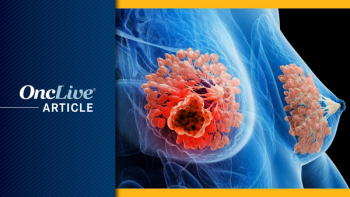
PRAME-directed therapies represent a novel approach in the treatment of advanced melanoma.
The combination of neoadjuvant vidutolimod and nivolumab elicited high response rates and was well tolerated in patients with high-risk, resectable melanoma, according to final results from a phase 2 trial.
The combination of neoadjuvant vidutolimod and nivolumab (Opdivo) elicited high response rates and was well tolerated in patients with high-risk, resectable melanoma, according to final results from a phase 2 trial (NCT03618641).
Data presented at the 2022 SITC Annual Meeting showed that 67% of patients who received the combination achieved any pathologic response (PR), defined as pathologic complete response (pCR) plus pathologic major response (pMR) and pathologic partial response (pPR). Specifically, 47% (n = 14), 10% (n = 3), 10% (n = 3), and 30% (n = 10) of patients achieved pCR, pMR, pPR, and pathologic non-response (pNR), respectively. Additionally, 57% of patients achieved major pathologic response, defined as pCR plus pMR.
“To evaluate the benefit of neoadjuvant intratumoral vidutolimod, we designed a phase 2 study to evaluate the effects of neoadjuvant intratumoral vidutolimod and nivolumab in high-risk resectable melanoma,” lead study author, Diwakar Davar, MD, of UPMC Hillman Cancer Center, and colleagues, wrote in a poster of the data.
The standard of care in high-risk, resectable melanoma is up-front surgery followed by adjuvant therapy with either PD-1 inhibitors for BRAF-mutant or wild-type disease, or dabrafenib (Tafinlar) plus trametinib (Mekinist) for BRAF-mutant disease. Neoadjuvant immunotherapy enhances systemic T-cell response to tumor antigens, which results in enhanced detection and killing of micrometastatic tumors that have disseminated beyond the resected tumor, which may be the cause of postsurgical relapse.
Vidutolimod, a virus-like particle (VLP), contains a CpG-A TLR9 agonist that activates plasmacytoid dendritic cells (pDCs) and stimulates type I interferon (IFN)–alpha production. Its polyguanylic and CpG motifs mimic retroviral viral DNA and induce systemic T-cell responses. CpG-A is the strongest known activator of tumor-associated pDCs for IFN and CFL induction, and its synthetic native DNA drives the strongest pDC response. CpG-A induces pDC differentiation into subsets distinct from those driven by other TLR9 agonists. Vidutolimod also has a lower inflammatory cytokine induction than other innate immune activators.
The second component of this immune-stimulating VLP is a Qbeta coat protein, which protects the G10 inside the VLP from in vivo degradation. This immunogenic protein induces a non-neutralizing antibody response after the first injection. The anti-Qbeta antibodies opsonize the VLP, facilitating its uptake into pDCs with enhanced induction of systemic CD8 T-cell response.
Patients were eligible to enroll in the phase 2 trial if they had stage IIIB, IIIC, or IIID cutaneous melanoma that was surgically resectable and had accessible tumors for biopsy and intratumoral vidutolimod.
The study enrolled 34 patients, 3 of whom withdrew consent after 1 dose of the study drug, and 1 of whom experienced disease progression after neoadjuvant therapy and was not eligible for resection.
In the preoperative neoadjuvant phase of this trial, patients received subcutaneous vidutolimod at 5 mg in week 1, then intratumoral vidutolimod at 10 mg once per week in weeks 2 through 7, plus nivolumab at 240 mg every 2 weeks, for a total of 7 weeks of treatment. Patients then underwent formal surgical reassessment and restaging scans, followed by definitive surgery. In the postoperative adjuvant phase of the trial, patients received 5 mg of subcutaneous vidutolimod plus 480 mg of nivolumab every 4 weeks for 48 weeks.
The primary end point was pCR per immune-related pathologic response criteria. Key secondary end points were safety, tolerability, relapse-free survival (RFS), distant metastasis–free survival (DMFS), and overall survival (OS). Pathologic responses were defined as pCR (0% residual viable tumor [RVT]), pMR (1%-10% RVT), PR (10%-50% RVT), and pNR (> 50% RVT).
The median age of 30 enrolled patients evaluable for efficacy was 61 years (range, 19-93), and 16 were male. One patient had received prior ipilimumab (Yervoy), and 1 had received a prior BRAF or MEK inhibitor. Regarding disease stage, 57% (n = 17), 37% (n = 11), and 6% (n = 2) had stages IIIB, IIIC, and IIID disease, respectively. Additionally, 5% (n = 17) had BRAF-mutant disease.
A total of 11, 15, and 4 patients had injected/measurable lesions in the head and neck, inguinal/axillary trunk, or extremities, respectively; 25 of these lesions were in the lymph nodes, and 5 were in transit.
Additional data showed The 1- and 2-year RFS rates were 94% and 88%, respectively, among patients who achieved any PR. Additionally, in those who achieved a PR, the 1- and 2-year DMFS rates were each 94%, and the 1- and 2-year OS rates were each 100%.
PR was associated with intra-tumoral accumulation of CD45+ and/or CD303+ cells, as well as significant increases in CD8+ T cells, CD4+ TFH cells, pDCs, and key signature scores of tumor lysis syndrome formation, and phagocytosis and macrophage activation.
When compared with an independent cohort of patients with melanoma treated only with a PD-1 inhibitor, a PR to neoadjuvant vidutolimod plus nivolumab was selectively associated with increased pDCs, with phagocytosis and macrophage activation demonstrating vidutolimod’s contribution to the observed activity. Additionally, pNR transcriptional analyses showed gene upregulation in IGF-1 and TGF-β, which was associated with non-response to PD-1 inhibitors.
In correlative analyses, PR was also associated with early, on-treatment increases in CD4+Ki67+ and CD8+Ki67+ T cells, which exhibited upregulation of activation and lytic activity markers. The investigators also noted targets for rational triplets in the form of LAG-3 upregulation on T cells in patients with pNRs. In addition, PR was associated with increased intermediate monocytes, increased pDCs, and reduced PD-L1 and PVR expression on pDCs.
The investigators performed a gut microbiota analysis at an intermediate post-vidutolimod, pre-surgery time point, where 24, 4, and 1 patients were evaluated on days 21, 49, and 7, respectively. In general, gram-negative rods at various time points before treatment and before or after surgery were consistently associated with PR. Specifically, Enterobacteriaceae and Oscillobacteriaceae spp. were associated with PR, and Lachnospiraceae spp. were associated with pNR.
Of the 34 patients evaluable for safety, the most common treatment-related adverse effects (TRAEs) included arthralgia/myalgia (grade 1, 22.6%; grade 2, 19.4%; grade 3, 3.2%), fever (grade 1, 45.2%; grade 2, 16.1%), flu-like symptoms (grade 1, 45.2%; grade 2, 25.8%), fatigue (grade 1, 45.2%; grade 2, 9.7%), a cytokine release syndrome–like reaction (grade 1, 6.5%; grade 2, 9.7%), colitis (grade 3, 3,2%), hypertension (grade 1, 6.4%; grade 2, 16.1%; grade 3, 9.7%), hyponatremia (grade 1, 61.3%), hypophosphatemia (grade 1, 38.7%; grade 2, 38.7%; grade 3, 3.2%), nausea/vomiting (grade 1, 12.9%; grade 2, 16.1%), anemia (grade 1, 29.0%; grade 2, 3.2%), thrombocytopenia (grade 1, 32.3%), injection site reaction (grade 1, 6.5%; grade 2, 12.9%), injection site infection (grade 1, 9.7%; grade 2, 9.7%; grade 3, 3.2%). No grade 4 or 5 TRAEs were observed.
“Future development of this combination is warranted,” the study authors concluded.
Davar D, Karunamurthy A, Chauvin J-M, et al. Neoadjuvant vidutolimod (vidu) and nivolumab (nivo) results in MPR and immune activation in high-risk resectable melanoma: final phase II clinical trial results. Presented at: 2022 SITC Annual Meeting; November 8-12, 2022; Boston, MA. Abstract 605