

|Videos|February 27, 2012
Dr. Erba Describes Proper Diagnostic Testing of CML
Author(s)Harry Erba, MD, PhD
Dr. Harry Erba, from the University of Michigan Health System, Describes Proper Diagnostic Testing of CML
Advertisement
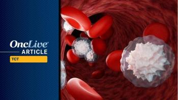
Harry Erba, MD, PhD, associate professor, Department of Internal Medicine, University of Michigan Health System, discusses the full histopathological examination that should be performed when a patient is diagnosed with chronic myeloid leukemia (CML).
If the routine complete blood count shows leukocytosis and a shift in maturity, basophilia, and splenomegaly are observed, 98 times out of 100 the patient will have CML, which is usually diagnosed in the chronic phase. This holds true whether constitutional symptoms are present or not.
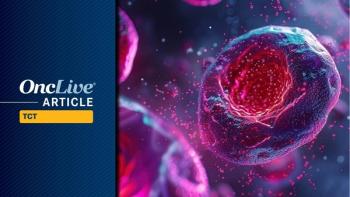
There are ways to make a non-invasive diagnosis for CML using fluorescence in situ hybridization (FISH) or polymerase chain reaction (PCR) to check for a BCR-ABL gene rearrangement in the peripheral blood. However, Erba does not recommend using this method, stating that CML requires a more thorough baseline analysis.
Bone marrow biopsy is critical at the time of diagnosis to provide morphologic evidence of the phase. The biopsy can show fibrosis or the clustering of mature cells indicating whether the cancer is in chronic, accelerated, or blast phase. Knowing the phase of the disease will help choose an appropriate therapy.
A cytogenetic analysis of the chromosomes at metaphase, the most condensed and easiest to distinguish phase in the cell cycle, may demonstrate clonal evolution or other changes that could upgrade the patient to a higher phase. The analysis may also show that some patients are not Philadelphia chromosome positive at metaphase, making it less likely to mistake a negative test later as a complete cytogenetic remission.
Additionally, Erba recommends the completion of a FISH analysis at the time of diagnosis. Approximately 10% of patients have a variant FISH pattern, which is important to know early on.
The final test that Erba recommends is a reverse transcription (RT)-PCR. The results of this test can be used as a baseline for response and to verify that the primers used in most commercial laboratories for detecting the BCR-ABL transcription are applicable. Some patients demonstrate variant break points, which makes it easier to arrive at the wrong conclusions if the RT-PCR test is not completed.
Advertisement
Related Content
Advertisement


Latest CME


Advertisement
Advertisement
Trending on OncLive
1
Nonresponse to Bridging Therapy and Peak ALC After Cilta-Cel Are Associated With Neurotoxicity, NRM in Myeloma
2
FDA Underscores Risks Associated With DPD Deficiency and Capecitabine/5-FU Use in Cancer Care
3
Long-Term Cilta-Cel Data Show Low Rates of PFS Events in Standard-Risk R/R Myeloma
4
Orca-T With Reduced-Intensity Conditioning Yields Robust Efficacy in Hematologic Malignancies
5