|Articles|July 9, 2020
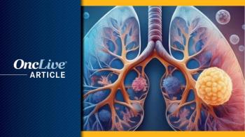
Investigators Leverage Radiomics to Risk Stratify Patients With Early-Stage Lung Cancer
Author(s)Caroline Seymour
Matthew Schabath, PhD, discusses the use of peritumoral and intratumoral radiomic features, where investigators created a model that identified patients with early-stage lung cancer at high risk of experiencing poor survival outcomes.
Advertisement
Through the use of peritumoral and intratumoral radiomic features, investigators created a model that identified patients with early-stage lung cancer at high risk of experiencing poor survival outcomes, according to new findings published in Nature Scientific Reports.1,2
The model, which evaluated the peritumoral feature Neighborhood Gray-Tone Difference Matrix (NGTDM) busyness and the intratumoral feature statistical root mean square (RMS), indicated that patients with early-stage, high-risk, screen-detected disease had worse overall survival (OS; HR, 9.91; P <.0001) versus patients with early low-risk disease (HR, 1.00). The 2.5- and 5-year survival rates were 25% and 0% versus 93% and 78%, respectively.
The model was also validated in an external cohort of patients with early-stage non-screen detected lung adenocarcinoma (n = 62). Here too, high-risk patients had worse OS (HR, 2.63; P = .112) versus low-risk patients (HR, 1.00). The 2.5- and 5-year survival rates were 56% and 42% versus 75% and 75%, respectively.
“Additional research is needed to inform us on the potential translational implications of this model, but it could make a major impact on saving lives by identifying lung cancer patients with aggressive disease while also sparring others from unnecessary therapy,” said study author, Matthew Schabath, PhD, associate member of the Cancer Epidemiology Department at Moffitt Cancer Center, in the news release.2
Predictive biomarkers that identify aggressive cancers from those that are indolent or carry a lower risk of poor prognosis represent an area of unmet need in the setting of lung cancer screening.
“Our goal was to use radiomic features to develop a reproducible model that can predict survival outcomes among patients who are diagnosed during a lung cancer screening,” said lead study author, Jaileene Pérez-Morales, PhD, of Moffitt Cancer Center, in the news release.
Radiomics is a growing field in which standard-of-care imaging is used to generate quantitative image features that can help inform risk prediction, diagnostic discrimination, prognostication, and treatment response. Previous studies have shown that peritumoral and intratumoral features carry prognostic and predictive information in lung, breast, gastric, and head and neck cancers. The noninvasive biomarker assessment methodology also compares favorably with circulating and tissue-based biomarker assays because the imaging captures the entire tumor burden rather than a sample of the tumor.
In the study, investigators compiled data from the National Lung Screening Trial and generated radiomic features from patients who had been diagnosed with lung cancer during their screening (n = 234), taking into account size, shape, volume, and textural intratumoral and peritumoral characteristics. The dataset was further divided into a training (n = 161) and a test cohort (n = 73).
A total of 109 peritumoral features and 155 intratumoral features were collected, of which 56 and 35 were found to be stable and reproducible, respectively.
In univariable analyses, 40 of the 91 radiomic features were significantly associated with OS in the training cohort. Thirty features were discarded because they were correlated. The 10 remaining features were refined to 4 using backward elimination. Among the four features, three were peritumoral (average co-occurrence joint entropy, NGTDM busyness, and average co-occurrence angular second moment) and one was intratumoral (statistical RMS). The four remaining radiomic features underwent Classification And Regression Tree analysis in the training cohort to create a working model.
The model, after refinement in the training cohort, stratified patients into 1 of 3 risk groups in the test cohort: low, intermediate, and high, the latter of which were deemed to have more aggressive cancer that, when caught early, might require frequent monitoring and/or adjuvant therapy.
Investigators found no statistically significant differences between the 3 risk groups by age, smoking status, number of pack-years smoked, self-reported chronic obstructive pulmonary disease, family history of lung cancer, histological subtypes, and treatment. However, statistically significant differences were observed across the 3 groups for sex (P = .04) and stage of disease (P = .001). Notably, 92% of the patients in the high-risk group were male versus 54% in the low-risk group (P = .04). Regarding disease stage, 33% of patients in the high-risk group had early-stage lung cancer versus 80% in the low-risk group (P = .001).
In the training cohort, the high-risk group was associated with poor OS (HR, 14.67; 10% 2.5-year OS and 0% 5-year OS; P <.0001) versus the intermediate- (HR, 3.25; 63% 2.5-year OS and 41% 5-year OS) and low-risk groups (HR, 1.00; 89% 2.5-year OS and 78% 5-year OS).
In the test cohort, the high-risk group was associated with poor OS (HR, 3.35; 50% 2.5-year OS and 0% 5-year OS; P = .043) versus the low-risk group (HR, 1.00; 68% 2.5-year OS and 51% 5-year OS). Similar findings were observed for progression-free survival (PFS).
Similar trends in survival were also observed after controlling for potential confounding variables in multivariable cox regression models. In the training cohort, the high-risk group was associated with worse OS (HR, 9.71; 95% CI, 3.85-24.48) and PFS (HR, 5.68; 95% CI, 2.32-13.93) compared with the intermediate- and low-risk groups.
The test cohort confirmed these findings with worse PFS in the high-risk group compared with the intermediate and low-risk groups (HR, 2.02; 95% CI, 0.34-11.99).
Among all patients, the high-risk group was associated with a significantly worse OS (HR, 5.16; 95% CI, 2.34-11.37) and PFS (HR, 4.02; 95% CI, 1.89-8.56) compared with the intermediate- and low-risk groups.
To determine the biological makeup of NGTDM busyness and statistical RMS, which were found to be the best indicators of peritumoral and intratumoral features, respectively, investigators evaluated a previously described dataset of patients with surgically resected lung adenocarcinoma (n = 103) who had available presurgical computerized tomography scans and gene expression data.
NGTDM is a texture feature that captures the intensity values of an array of pixels, through coarseness, contrast, and busyness, to identify the difference between the centermost element of the environment evaluated. Statistical RMS is an intensity feature that is used to determine the RMS of the centermost element’s intensity value.
NGTDM busyness was found to be significantly associated with RABGAP1L, where high expression was more commonly observed in intermediate- and high-risk patients. Additionally, RMS was significantly associated with FOXF2 (r = .45) and LOC285043 expression (r = .44). Intermediate- and high-risk patients had lower expression of FOXF2 versus low-risk patients, whereby the authors concluded that low expression thereof could be a negative prognostic indicator.
References:
- Pérez-Morales, J, Tunali, I, Stringfield, O, et al. Peritumoral and intratumoral radiomic features predict survival outcomes among patients diagnosed in lung cancer screening. Nature Sci Rep. 2020;10(10528). doi:10.1038/s41598-020-67378-8
- Moffitt researchers develop tool to detect patients at high risk for poor lung cancer outcomes [news release]. Moffit; July 1, 2020. Accessed July 7, 2020. https://bit.ly/3iIuSCD
Advertisement
Related Content
Advertisement


Advertisement
Advertisement
Trending on OncLive
1
FDA Grants Priority Review to Giredestrant for ER-Positive Early Breast Cancer
2
Bispecific-Based Approaches Highlight Key Multiple Myeloma Data Set to Emerge at EHA 2026
3
HERTHENA-Breast04 Is Set to Delineate the Role of HER3-Targeted Therapy in Advanced HR+ Breast Cancer
4
First-Line Zanubrutinib Is Associated With Improved Real-World Efficacy in Older Patients With CLL
5