Nirav N. Shah, MD, MSHP, discusses bexobrutideg and what the BTK degrader could bring to chronic lymphocytic leukemia management.
Antibody-drug conjugates represent a targeted class of agents with an improved therapeutic index over traditional chemotherapy in the treatment of non-Hodgkin lymphoma.
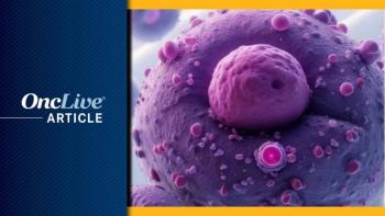
Antibody-drug conjugates (ADCs) represent a targeted class of agents with an improved therapeutic index over traditional chemotherapy in the treatment of non-Hodgkin lymphoma (NHL). Despite the potential of these agents, the delivery and release of cytotoxic agents to malignant cells through a combination of monoclonal antibody, payload, and linker represent a complex challenge. Nevertheless, because of recent innovations in the treatment of B-cell malignancies, the number of ADCs in clinical trials has increased in recent years. Moreover, these pipeline agents, such as polatuzumab vedotin, offer the potential to change the treatment landscape for NHL and other hematological malignancies through improved targeting and greater potency, compared with early-generation ADCs. This supplement reviews the development of ADCs, assesses the potential of investigational agents, and evaluates their role in the evolving treatment spectrum for lymphoma.Non-Hodgkin lymphoma (NHL) comprises a heterogeneous group of lymphoid malignancies that originate in B cells, T cells, or natural killer cells and exhibit distinct clinical behavior and variable responses to treatments.1-3 NHL is the sixth most common cause of cancer death in the United States.4 In 2018, it is estimated that 74,680 new cases and 19,910 deaths due to NHL will occur in the United States.4 NHL is most prevalent in patients ≥70 years and in men.4
Mature B-cell neoplasms constitute 80% to 85% of NHLs.5 The most prevalent B-cell lymphomas are diffuse large B-cell lymphoma (DLBCL) and follicular lymphoma (FL), together representing approximately 65% of all NHL cases.5 NHL can be indolent or aggressive.6 Accounting for between 30% and 40% of NHL cases, DLBCL, an aggressive NHL subtype with multiple variants, represents the most common histologic subtype6,7 and predominantly affects adults.8 In 2016, it was estimated that 27,650 new cases of DLBCL would be diagnosed in the United States.8 Despite improvements in first-line therapy during the past decade, up to 40% of patients with DLBCL will develop relapsed or refractory (R/R) disease, representing a significant cause of morbidity and mortality.6,9
FL is the second most common form of NHL, comprising one-fifth of all NHL cases and accounting for up to 70% of the indolent lymphomas reported in clinical trials in the United States and Europe.3 Although FL usually follows an indolent course, it can lead to poor outcomes with relapse occurring within 2 years of initial treatment.6Treatment of NHL is based on the patient's histologic subtype, extent of disease, and age.2 In addition, most chemotherapy agents target rapidly proliferating cancer cells nonspecifically, and they have toxic effects on normal tissue.7 Monoclonal antibodies, which usually offer inadequate clinical activity when used as monotherapy, are often used in conjunction with chemotherapy to treat lymphoma.7 First approved in the United States in 1997,10 rituximab is an anti-CD20 monoclonal antibody that has transformed the therapeutic landscape of B-cell malignancies, marking a paradigm shift with the beginning of targeted agents to treat lymphoma.7,10 Other monoclonal antibodies, such as obinutuzumab, a newer glycoengineered type II anti-CD20 monoclonal antibody, have also been incorporated into the treatment armamentarium for lymphoma.2
ADC indicates antibody—drug conjugate; DM1, DM4, derivatives of maytansine; MMAE, monomethyl auristatin E. Reprinted from Panowski S, Bhakta S, Raab H, Polakis P, et al. Site-specific antibody drug conjugates for cancer therapy. MAbs. 2014;6(1):34-45.
First- and second-line treatment options for patients with FL vary according to age and include chemotherapy and chemoimmunotherapy combinations or single-agent therapies.2 First-line treatment options for patients with DLBCL typically include anthracycline-based combination chemotherapy regimens comprising cyclophosphamide, doxorubicin, vincristine, and prednisone (CHOP) in combination with the anti-CD20 monoclonal antibody rituximab (R-CHOP).2,6 Second-line treatment options for patients with DLBCL are stratified according to suitability for high-dose therapy using other chemotherapy combinations with or without rituximab or single-agent therapies.2
Chemotherapy is associated with complete response (CR) rates between 40% and 50% in patients with NHL.6 Many patients who initially respond to chemotherapy will eventually relapse, with rates of overall survival (OS) at 3 years reaching approximately 30%.11 Despite recent therapeutic advances in lymphoid malignancies, treatment challenges remain in the management of DLBCL and other R/R NHLs.6Several monoclonal antibodies (eg, bevacizumab, rituximab, and cetuximab) are used in standard treatment regimens for solid tumors and hematological cancers.12 The use of traditional chemotherapies (eg, vinblastine, doxorubicin, and paclitaxel) for cancer treatment is limited due to the nonspecific toxicities of these drugs, resulting in a narrow therapeutic window and increased drug resistance.13 The development of antibody-drug conjugates (ADCs) serves to bridge the gap between the antibody and cytotoxic drug, leading to the introduction of targeted agents with an improved therapeutic index. 13,14 An ADC consists of a monoclonal antibody conjugated to a cytotoxic agent via a linker.15 The design of ADCs enables the delivery and release of cytotoxins directly to malignant cells, providing high specificity in anticancer activity.7
The careful selection of the 3 ADC components (monoclonal antibody, cytotoxic agent, and linker) enables the development of safe, efficacious conjugated therapies. 16 Each component contained within an ADC will be discussed here in detail (Figure 1).17ADCs use high-affinity monoclonal antibodies to deliver and release cytotoxic agents at the tumor site.16 The intravenous administration of ADCs prevents the degradation of the monoclonal antibody by gastric acids and proteolytic enzymes.16 The monoclonal antibody enables the circulation of the ADC in the bloodstream where it binds to the target antigen on the tumor cell surface,16 reducing the likelihood of systemic exposure and therefore limiting toxicity.16 The monoclonal antibody should be specific for the surface antigen on malignant cells to enable the accumulation of the cytotoxic drug at the tumor site and to ensure the targeted delivery of the high-potency cytotoxic agent.15,16The cytotoxic agent (or payload) impedes cellular mechanisms necessary for tumor survival by disrupting microtubule assembly or inducing DNA damage, causing cell death directly or through apoptosis.16 The cytotoxic agent should be highly potent, with IC50 (ie, the concentration needed to achieve 50% inhibition) values in the nanomolar or picomolar range.18 If it is assumed that all steps involved in the mechanism of ADC action have 50% efficiency, then only 1% to 2% of the administered drug is expected to reach tumor cells.19 Therefore, selecting a cytotoxin that is highly efficacious at very low concentrations is preferable and drugs that would not normally be considered for chemotherapy due to excessive toxicity have a useful role in ADCs.16Linkers serve as short chemical spacers that bind the drug to the antibody, which must be stable in circulation. 19 The properties of the linker influence the therapeutic index, efficacy, and pharmacokinetics of the ADC.13 Stable linkers in ADCs can maintain the antibody concentration in the blood circulation and do not release the cytotoxic drug before reaching the target, limiting nontarget toxicity. At the same time, linkers must be labile enough to allow the efficient release of the cytotoxic agent once the ADC is internalized within the tumor cells.19Important considerations in the design of ADCs are target antigen selection, target internalization, linker stability, conjugation, and cytotoxic payload,15 and will be discussed here.An essential function of ADCs is to promote the efficient internalization of the cytotoxic drug within malignant cells while minimizing toxic effects on healthy cells.16 This function relies on the effective binding of the monoclonal antibody component to the target antigen, which should be tumor specific or tumor associated.16 The monoclonal antibody should display high immuno-affinity for the target antigen.16 In addition, the target antigen should display high penetrance and high expression at the tumor site, with limited expression on healthy tissues to reduce any adverse effects.7,13,16Before the cytotoxic drug can be released in its active form, the ADC-target complex must internalize into the target cells via receptor-mediated endocytosis.16 The efficiency of internalization affects drug uptake and release,15 and it is determined by the target itself and the choice of cytotoxin.16 Targets may internalize irrespective of ligand binding, or they may reside permanently on the cell surface. Barriers to reaching the threshold concentration of the cytotoxic drug include inefficient internalization and limited target antigens.16 In addition, the specific epitope that binds to the monoclonal antibody may influence the level of antigen-antibody affinity and play a role in determining internalization efficiency.20Linkers can be modified to impact the biophysical and functional performance in a positive or negative manner by affecting the safety, specificity, potency, and activity of ADCs.17 The stability of the linker has a considerable influence on the toxicities that are exerted by the payload.13 The linker should be stable in circulation and must be labile enough to efficiently release the cytotoxic agent inside the tumor cell.19 There are some linkers that remain stable in the cell, and degradation of the antibody and linker are necessary to release the cytotoxic agent.15 The nature of ADC-associated toxicities is affected by the stability of the linker. Linkers that are more stable behave in a target-specific manner to release their payload and tend to result in specific toxicities; conversely, less stable linkers, which are likely to undergo nonspecific cleavage, tend to have a broader toxicity profile.21
Linkers are generally classified into 2 categories: cleavable linkers, which encompass hydrazone, disulfide, and peptide linkers, and noncleavable linkers, which require degradation by lysosomes after internalization.16 Hydrazone linkers are acid sensitive and take advantage of the low pH (4-6) within the endosomes and lysosomes of cancer cells, undergo acid hydrolysis, and release the cytotoxic payload.22,23 Disulfide linkers are glutathione sensitive and more stable in the circulation. These linkers exploit higher concentrations of glutathione, which are present in cancer and associated with cell survival and tumor growth.13,23 Peptide linkers are protease sensitive and more stable than the other forms of cleavable linker. These linkers are designed to release the payload upon cleavage by proteases and enable improved control of cytotoxic drug release.23
Different mechanisms are utilized to cleave these various linkers from the payload in the cell. These include acidic degradation (hydrazones), thiol-disulfide exchange reactions (disulfide, carbonate), and protease cleavage by cathepsin B (dipeptide).21 Noncleavable linkers (eg, thioether or maleimidocaproyl) use lysosomal enzymes to break down the ADC and release the cytotoxic payload after internalizing at the tumor site.13 For example, ado-trastuzumab emtansine (trastuzumab) utilizes a noncleavable maleimidocaproyl linker that links the maytansinoid toxin to the HER2 antibody.24 This design allows trastuzumab to remain stable in serum for more than 3 days.25
Two conjugation approaches are typically used to attach linkers to antibodies.26 The first approach results in heterogeneous payload placement and exploits the reactivity of endogenous lysine or hinge cysteine residues within the antibody. The second approach results in site-specific payload placement and uses reactive chemical handles that are exogenously introduced into particular locations on the antibody through various means. Site-specific conjugation techniques are continuing to evolve, and the functional and analytical advantages of employing site-specific conjugation are being recognized. Nevertheless, heterogeneous conjugates still dominate the ADC pipeline.18An important attribute to consider for any ADC is the amount of drug loading, or the average ratio of conjugated payload to antibody, known as the drug-to-antibody ratio (DAR). This ratio determines the amount of payload per antibody delivered upon internalization and, in turn, affects both efficacy and toxicity.18 Optimizing the DAR is necessary because low drug loading will lead to lower efficacy, whereas high drug loading can negatively affect pharmacokinetics and further increase instability and toxicity.27
Different conjugation methods involved in the synthesis of ADCs result in a heterogeneous population of constructs with multiple DARs.21 As such, many current ADCs are characterized by heterogeneous payload conjugation to lysine or cysteine residues. A mixture of ADC species results from the random nature of both conjugation approaches, with DARs ranging from 0 to 8.16,26 These variations in the DAR can have substantial effects on the disposition of an ADC (eg, tendency to aggregate systemically, efficacy of endocytosis, half-life, and ability to bind target antigens),28,29 with implications for toxicity.21
Greater toxicity is associated with the more highly conjugated species, which is thought to be caused by increased hydrophobicity and faster clearance by the liver.26 The heterogeneity found in ADCs results in mixtures of inactive (unconjugated) and more toxic isoforms and lowers the therapeutic index.26
Extensive research has supported a DAR of approximately 4 as an optimal number of drug molecules per antibody to maximize the efficacy of most ADCs.28 This ratio is generally favored as it reduces the amount of unconjugated antibody, limits the circulation half-life to that of the unmodified antibody, and does not significantly affect antigen binding.7The potency of the cytotoxic payload is a key consideration in the design of ADCs. Highly potent cytotoxic drugs are used as payloads in ADCs and exert their effects on critical cellular processes required for survival. Most ADCs under investigation utilize either maytansine derivatives (DM1/DM4) or auristatins (monomethyl auristatin E and F [MMAE/MMAF]), which are both microtubule inhibitors that are cytotoxic to slowing replicating or nondividing tumor cells. These agents typically induce apoptosis in cells undergoing mitosis by causing cell cycle arrest at G2/M. It has been demonstrated that microtubule inhibitors may also disrupt nondividing cells in interphase.30 Other cytotoxic drugs used in ADCs include enediynes (calicheamicin), duocarmycin derivatives, pyrrolobenzodiazepines, and indolinobenzodiazepines, all of which target the minor groove of DNA, and quinoline alkaloids, which inhibit topoisomerase I. Only a minute amount (<1%) of the injected dose of antibody localizes to the tumors, so cytotoxic payloads must be highly potent, often with IC50 potency values in the picomolar range.31 This potency determines the toxic qualities of ADCs, resulting in the majority of toxicities being characterized by the class of payload.21Early-generation ADCs were constrained by several limitations related to immunogenicity, potency, target selection, and tumor selectivity.15 Limitations of Early-Generation ADCs Immunogenicity. First-generation ADCs consisted of murine monoclonal antibodies covalently linked to anticancer drugs (eg, doxorubicin, methotrexate, vinblastine).15 Murine antibodies caused severe immunogenicity by evoking an immune response and producing human antimouse antibodies in patients.13,32,33 The immune response evoked by murine antibodies lowers the effectiveness of the ADC and may occur rapidly, resulting in the formation of human anti-mouse antibodies within 2 weeks of administration.34
Early cytotoxins such as doxorubicin (an anthracycline) or methotrexate (an antimetabolite/antifolate) were utilized as payloads in first-generation ADCs.35,36 These cytotoxins have established efficacy when used as standard chemotherapy; however, they are known to have insufficient potency at low concentrations when conjugated to a monoclonal antibody.37
Target selection. Lack of specificity to the target antigen in early-generation ADCs contributed to their cytotoxic effects on healthy tissue.16 Other characteristics of the ADC target antigen should be considered, such as poor internalization or inadequate antigen binding.16
The differences in target antigen requirements for cleavable versus noncleavable linkers suggest that the internalization and trafficking component of ADC processing plays a critical role in its function.38 Cleavable linkers are sensitive to the microenvironment in endosomal compartments, with the trafficking requirements for the release of attached payloads thought to be less stringent than those for ADCs carrying noncleavable linkers, which require complete lysosomal degradation before payload release.16 The nature of the overall relationship between internalization, intracellular trafficking, and ADC potency remains poorly understood.39Based on monoclonal antibodies that did not undergo internalization, early-generation ADCs were engineered to release the cytotoxic payload upon cleavage of the linker by tumor-specific enzymes (such as matrix metalloproteinases) or in tumor-specific environments (such as a lower pH).40 These ADCs failed to undergo internalization and did not improve drug specificity or lower toxicity.The concept of combining a monoclonal antibody with a cytotoxic drug to form an ADC has existed for more than 4 decades,27 and substantial improvements have been made since the first generation of ADCs.15 New-generation ADCs have several features that are designed to address the limitations of earlier ADCs.15Bioengineering research has enabled the discovery of chimeric, humanized, and fully human antibodies, which have replaced the murine antibodies used in early-generation ADCs.14 These newer antibodies have decreased immunogenicity compared with their earlier counterparts and offer other advantages over murine antibodies, including longer circulation and improved antibodydependent cellular cytotoxicity.41The cytotoxins employed by newer-generation ADCs are considerably more potent than the anticancer agents used in the past (eg, doxorubicin and methotrexate).16 Cytotoxic agents used in ADCs under investigation for the treatment of patients with NHL are discussed here and mainly fall into 2 classes: microtubule inhibitors (DM1, DM4, MMAE, MMAF) or DNA synthesis inhibitors (calicheamicins, duocarmycins, pyrrolobenzodiazepine dimers, and doxorubicin).18 Other cytotoxic agents have been utilized in ADCs with novel mechanisms of action (eg, irinotecan derivatives and alpha-amanitin).18
Dolastatin 10 and maytansine are potent antimitotic microtubule-disrupting agents with IC50 potency values in the nanomolar range. DNA-alkylating agents have IC50 potency values in the picomolar range.18,19 The largest group of ADCs in clinical trials are MMAE and MMAF, which are derivatives of dolastatin 10. Dolastatin 10 failed a phase II study in patients with prostate cancer due to lack of efficacy.42 Auristatins are antimitotic cytotoxins that strongly bind tubulin and inhibit polymerization, leading to cell cycle arrest.43 MMAE and MMAF have been singled out for further study due to several desirable characteristics, including their physiologic stability, potency, aqueous solubility, and ease of attachment of stable linkers.15,44
Clinical trials of maytansine were conducted in the 1970s and later discontinued due to a high level of systemic toxicity.42 Derivatives of maytansine, however, are highly potent cytotoxic agents when linked to a targeting antibody and comprise the largest group of ADCs behind auristatins currently in clinical trials.44
Second-generation maytansinoids include DM1 and DM4, which are cytotoxic agents used with trastuzumab and coltuximab, respectively.15,45
Calicheamicins are highly potent enediyne antitumor antibiotics that cleave DNA with high specificity by binding in the DNA minor groove and inducing double-strand breaks.43,47 Gemtuzumab and inotuzumab utilize calicheamicin payloads, which are associated with narrow therapeutic indices and serious late toxic effects.47 Due to its hydrophobic interactions, only a few molecules of calicheamicin per immunoglobulin can be conjugated before high levels of aggregated protein appear.45
Similar to calicheamicins, duocarmycins are potent antitumor antibiotics that are DNA minor groove-alkylating agents. A phase I study investigated BMS‑936561 (also known as MDX‑1203), a conjugate of a human anti‑CD70 antibody (MDX‑1115), and a duocarmycin analogue (MED‑2460), in patients with advanced clearcell renal cell carcinoma and B‑cell NHL. BMS‑936561 was shown to be well tolerated at doses of up to 8 mg/kg.45
Pyrrolobenzodiazepines, which are naturally occurring antitumor antibiotics, bind to the DNA minor groove in a sequence-specific manner.43 Pyrrolobenzodiazepine dimers are composed of 2 pyrrolobenzodiazepine units that use different tethers to yield symmetrical and nonsymmetrical dimers. This configuration allows the resulting compound to crosslink DNA by binding to the N2 position of guanine on opposing strands of DNA.45,47
Other approaches are being explored to enhance the potency of ADCs; these include strategies to reduce resistance against the cytotoxin or to increase tumor penetration by using of smaller protein scaffolds.44 In addition to demonstrating efficacy as monotherapy, ADCs have shown synergy when used in conjunction with monoclonal antibodies, including checkpoint inhibitors.15,44Another important consideration in ADC design is the careful selection of an appropriate target antigen and antibody. Notably, the antibody should bind accurately and efficiently to the antigen on tumor cells while sparing nonmalignant cells. Selective expression of the target antigen on tumor cells and internalization upon antibody engagement are required for successful ADC approaches.17,21
The effectiveness of the ADC may be limited by insufficient antigen expression on tumor cells, inappropriate or low-affinity binding, or a lack of internalization following binding.21 To ensure that adequate amounts of the cytotoxic drug are delivered by the ADC, an adequate level of antigen expression is necessary. The level of antigen expression needed for efficient ADC activity depends on the specific antigen in question.16 A minimum threshold of approximately 10,000 antigens per cell has been identified for the delivery of lethal quantities of the cytotoxic drug.16,48 In B-cell lymphoma, the level of CD19 antigen expression may be limited to as few as 30,000 per cell to elicit ADC activity.16,49The monoclonal antibody component of the ADC binds to the target antigen and should retain high immunoaffinity, typically in the 0.1- to 1-nanomolar range.13,16 The internalization process takes place via receptor-mediated endocytosis, which is completed by the formation of a clathrin-coated early endosome containing the ADC antigen complex.16,49 Internalization must be efficient enough to ensure that the necessary threshold concentration of the cytotoxic agent is reached given the level of antigen expression within the cell.16 In addition to antigen affinity, other important properties of antibodies used in ADCs include target specificity, good retention, minimal immunogenicity, low cross-reactivity, and a long circulation in plasma.13
The process of internalization and subsequent trafficking through endosomal and lysomal compartments has been shown to vary among target antigens.37 Polson et al studied the in vivo efficacy of ADCs against 7 NHL target antigens and found that ADCs bearing cleavable linkers could mediate tumor growth inhibition against all 7 targets, whereas ADCs bearing noncleavable linkers were efficacious against only 2 of the 7 targets. Thus, the choice of linker is a key consideration that can impact selectivity and internalization of the ADC.
Site-specific conjugation has been used to yield ADCs with higher DARs, specifically between 6 and 8.50 This method may be useful for overcoming low-expression target antigens and slower tumor cell internalization kinetics, while achieving a high therapeutic index.50 However, a higher DAR is associated with deterioration of certain ADC attributes, such as increased systemic clearance, reduction in therapeutic efficacy, lower stability under stressed conditions,51 heterogeneity, and higher propensity to aggregation, presumably due to the hydrophobic nature of the payloads.52 Adverse clinical effects, including immunogenicity, have been associated with protein aggregation.53
There has been much interest in exploring how sitespecific conjugation techniques can be used to alter the pharmacokinetics of ADCs.21 Site-specific conjugation techniques currently under investigation include the use of engineered cysteines, unnatural amino acids, and the addition of a selenocysteine, glutamine, or aldehyde tag.30 These technologies can achieve a more homogeneous population of ADCs by engineering conjugates with specific DARs.21 This may result in an improved therapeutic window with enhanced efficacy and decreased toxicity.Four ADCs are approved in the United States: a CD33- targeting agent indicated for acute myeloid leukemia, an HER2-targeting agent indicated for HER2-positive metastatic breast cancer, and 2 agents indicated for lymphoid malignancies that target CD30 and CD22.17 In addition, several ADCs targeting other B-cell surface antigens are in development for the treatment of NHL (Table).54Initially approved in the United States in 2000 for the treatment of acute myeloid leukemia, gemtuzumab was withdrawn in 2010 due to concerns of treatment-related toxicity.17,55,56 In 2017, gemtuzumab was reapproved with a lower recommended dose and altered dosing schedule. Gemtuzumab is a humanized anti-CD33 antibody attached covalently to the cytotoxic agent N-acetyl-gamma-calicheamicin via an acid-labile hydrazone bifunctional linker.56,57Trastuzumab was approved in the United States in 2013 for treating HER2-positive metastatic breast cancer and is the first ADC approved for a solid tumor indication.17,57,58 Trastuzumab is a humanized anti-HER2 antibody attached covalently to the microtubule inhibitor DM1 (a maytansine derivative) via the stable thioether linker MCC (4-[N-maleimidomethyl] cyclohexane-1-carboxylate).58CD30-Targeting Agent. Brentuximab vedotin (brentuximab; SGN-35) was initially approved in the United States in 2011, and it now has multiple indications in the treatment of non-Hodgkin lymphoma and anaplastic large-cell lymphoma.59 Brentuximab is a chimeric anti- CD30 monoclonal antibody attached covalently to the synthetic microtubule disrupting agent MMAE via a cathepsin B-sensitive cleavable dipeptide valine-citrulline linker.13,16,60 The CD30 antigen is a transmembrane protein that belongs to the tumor necrosis factor receptor family and is expressed in several lymphoma subtypes, including anaplastic large cell lymphoma and Hodgkin lymphoma, which is characterized by CD30-positive Reed-Sternberg cells.61,62 Upon attachment to the receptor, brentuximab is internalized by CD30-expressing cells. Cathepsin B is a tumor-specific protease that recognizes and cleaves the valine-citrulline dipeptide bond inside the tumor cells.13,16
ADC indicates antibody—drug conjugate; BCR, B-cell receptor; DLBCL, diffuse large B-cell lymphoma; FL, follicular lymphoma; NHL, non-Hodgkin lymphoma; SEA, sugar-engineered antibody; vcMMAE, valine–citruline monomethyl auristatin E.
Reprinted from: Blood Reviews. Ku M, Chong G, Hawkes EA. Tumour cell surface antigen targeted therapies in B-cell lymphomas: Beyond rituximab. Vol 31, No 1, Page 24 [Table 1]. Copyright Elsevier 2017.
This action releases MMAE into the cytosol where it inhibits microtubule formation and results in apoptosis.62 Brentuximab has been investigated in phase II studies for the treatment of NHL, including DLBCL. One such study evaluated the safety and efficacy of brentuximab plus R-CHOP in the frontline treatment of patients with high-intermediate or high-risk DLBCL (ClinicalTrials. gov identifier: NCT01925612).63 Of the 51 patients who received the study treatment, 32 were high-intermediate risk (International Prognostic Index [IPI] score of 3, ageadjusted IPI score of 2), and 19 were high risk (IPI of 4 to 5, age-adjusted IPI of 3).63 The overall response rate (ORR) as measured by PET at the end of treatment was 97% (80% CR).63 The rate of CR was 92% in patients who were CD30 positive (n = 13). Four patients (all CD30 negative) had progressed after a median follow-up of 5 months.63 Activated B-cell-like and germinal center B-cell-like subtypes showed similar CR rates.63
The most frequent treatment-emergent adverse events (AEs) were nausea, fatigue, peripheral sensory neuropathy, diarrhea, anemia, decreased appetite, febrile neutropenia, and vomiting.63 The most common grade ≥3 AEs were febrile neutropenia (27%), neutropenia (25%), and anemia (24%).63 Neuropathy (mostly grade 1-2) was reported in 55% (38%, 1.2 mg/kg brentuximab plus R-CHOP; 17%, 1.8 mg/ kg brentuximab plus R-CHOP). Treatment discontinuation due to AEs occurred in 10% of patients.63
A phase II, open-label, multicenter study evaluated brentuximab in patients with both CD30-expressing and CD30-nonexpressing R/R NHL (ClinicalTrials.gov identifier: NCT01421667).64 The study included 68 patients with CD30-positive (≥1%) B-cell NHL (mostly DLBCL, 49/68).64
The ORR was 44% (17% CR) in patients with DLBCL (median duration of response [DOR], 5.6 months) and 26% (16% CR) in those with B-cell NHL.64 In the DLBCL cohort, the median DOR was 16.6 months in patients who achieved CR.64 The rate of CR in patients with DLBCL was 17% in those who were CD30-positive and 10% in those who were CD30-negative.64 Stable disease was maintained in 23% (n = 11), and 33% (n = 16) experienced disease progression.64 No significant correlation was observed between response and CD30 expression level.64
The most common treatment-emergent AEs (all grades) in patients with DLBCL included fatigue (55%), diarrhea (43%), neutropenia (41%), nausea (39%), decreased appetite (31%), pyrexia (31%), and peripheral sensory neuropathy (29%).64 Treatment discontinuation due to AEs occurred in 12% of patients with DLBCL.64Inotuzumab ozogamicin (inotuzumab; CMC544) was approved in 2017 in the United States for the treatment of R/R B-cell precursor acute lymphoblastic leukemia.65 Inotuzumab is a humanized anti-CD22 monoclonal antibody attached covalently to N-acetyl-gamma-calicheamicin via a cleavable hydrazone linker.18,57,65 The CD22 antigen is a sialic acid-binding immunoglobulin-like lectin that is expressed in most (>90%) patients with DLBCL and FL.57
A phase I study evaluated inotuzumab in patients with relapsed CD22-positive B-cell NHL (n = 79).66 The median age was 60 years.66 The maximum tolerated dose (MTD) was determined to be 1.8 mg/m2 every 3 weeks.66 The ORR was 68% (32% CR/unconfirmed CR [CRu]) in patients with FL and 15% (7.7% CR/CRu) in patients with DLBCL.66 Median progression-free survival (PFS) was 317 days in patients with FL and 49 days in patients with DLBCL.
Median OS was 193 days in patients with DLBCL and had not yet been reached in patients with FL.66 In all patients receiving the MTD, the most common AEs (all grades) were thrombocytopenia (91%), asthenia (67%), and nausea and neutropenia (51% each).66 Treatment discontinuation due to thrombocytopenia occurred in 22% of patients.66
A phase I/II study included a dose-escalation stage to determine the MTD of the inotuzumab-rituximab combination in patients with relapsed FL and DLBCL followed by an expanded cohort to further evaluate the safety and efficacy at the MTD in 3 groups of patients: relapsed FL, relapsed DLBCL, and refractory NHL (ClinicalTrials. gov identifier: NCT00299494).67,68 The median age in the overall study population was 65 years.73 The MTD was 1.8 mg/m2 for the expanded cohort (n = 104).68 The ORR was 87% (62% CR) in patients with relapsed FL and 74% (50% CR) in patients with relapsed DLBCL.68 In patients with relapsed FL, the 2-year PFS and OS rates were 68% and 91%, respectively.68 In patients with relapsed DLBCL, the 2-year rates of PFS and OS were 41% and 69%, respectively.68
The most common AEs (all grades) were thrombocytopenia (56%), nausea (57%), fatigue (53%), increased aspartate transaminase (41%), neutropenia (33%), increased alkaline phosphatase (30%), and vomiting (28%).68 The most common grade 3/4 AEs were thrombocytopenia (9%) and neutropenia (8%).68 Treatment discontinuation due to AEs occurred in 49% of patients.68 A phase 3 randomized, open-label, 2-arm study (B1931008) compared the safety and efficacy of combined inotuzumab and rituximab therapy with the investigator's choice in patients with R/R aggressive NHL (ClinicalTrials.gov identifier: NCT01232556).69.70 Despite reports of clinical activity in earlier phase I/II studies, results from an interim analysis showed that inotuzumab would not meet the primary objective of improving OS; no new safety issues were identified.70,71Coltuximab ravtansine (coltuximab; SAR3419) is a humanized anti-CD19 monoclonal antibody conjugated to the potent antimitotic maytansinoid derivative DM4 via a cleavable disulfide N-succinimidyl-4-(2-pyridyldithio) butyrate linker.18,54 The CD19 antigen is a transmembrane glycoprotein that plays a role in the regulation of B-cell receptor signaling, is expressed on the surface of mature B cells, and is quickly internalized following antigen binding.7
Two phase I studies evaluated coltuximab in patients with R/R FL and DLBCL.72,73 One of these, an open-label, dose-escalation study, explored the MTD of coltuximab in patients with R/R B-cell NHL (ClinicalTrials.gov identifier: NCT00539682).72,74 Twenty-six of 35 evaluable patients with R/R B-cell NHL showed tumor reduction (74%) as measured by PET-CT.72 In patients with rituximab- refractory disease (n = 15), 47% (7) patients showed tumor reduction.72 The MTD was established at 160 mg/m2; the ORR was 24% in the 17 evaluable patients at MTD.72 Grade 3/4 AEs included eye disorders (15%), neutropenia (10%), peripheral neuropathy (8%), and dyspnea (3%).72
A different phase I dose-escalation study in 69 patients with R/R CD19-positive B-cell NHL explored the recommended dose of coltuximab, which was determined to be 55 mg/m2.73 Due to the occurrence of late or cumulative toxicities, the dosing schedule was changed from 8 to 12 weekly doses to 4 weekly doses, followed by 4 biweekly doses, resulting in an improved safety profile.73 With both dosing regimens, the ORR was 30% in patients with indolent FL and DLBCL.73
Two separate phase II studies evaluated coltuximab as monotherapy and in combination with rituximab in patients with R/R DLBCL. STARLYTE was a phase II study that evaluated single-agent coltuximab in patients with CD19-positive R/R DLBCL who were ineligible for transplantation after at least 1 standard rituximab-based therapy (ClinicalTrials.gov identifier: NCT01472887).75,76
Coltuximab 55 mg/m² was administered weekly for 4 weeks, then biweekly until disease progression or other study discontinuation criteria (N = 41).76 The median age was 71 years. 76 The ORR was 44% (12% CR). OS and PFS were unavailable due to inadequate follow-up.76 The most common AEs (all grades) included nausea (23%), diarrhea (20%), fatigue (18%), coughing (18%), vomiting (13%), decreased appetite (13%), asthenia (12%), abdominal pain (12%) and back pain (12%).76 Grade 3/4 hematological toxicities included neutropenia (26%), thrombocytopenia (10%), and anemia (7%).76 Treatment discontinuation due to AEs occurred in 4 patients.76
A different phase II, open-label, single-arm study evaluated coltuximab plus rituximab in patients with R/R DLBCL.77 A total of 52 patients with R/R DLBCL received 4 doses of 55 mg/m2 coltuximab with 375 mg/ m2 rituximab, first weekly and then every 2 weeks for an additional 8 weeks (N = 45) in the per-protocol population.77 The median age was 67 years.77 The ORR was 58% in relapsed patients and 31% in refractory patients; also, the objective response (OR) was 43% in patients refractory to their previous chemotherapy regimen and 15.4% in patients refractory to first-line therapy.77 The most common AEs (all grades) included gastrointestinal disorders (52%) and asthenia (25%). No treatment discontinuation occurred due to AEs.77
Denintuzumab mafodotin (denintuzumab; SGN-CD19A) is a humanized anti-CD19 monoclonal antibody conjugated to the auristatin MMAF via a noncleavable maleimidocaproyl linker.18 A phase I, multicenter, openlabel, dose-escalation study evaluated the safety and tolerability of denintuzumab in patients with R/R B-cell NHL (ClinicalTrials.gov identifier: NCT01786135).78,79 The median number of prior therapies was 2 and the median patient age was 65 years; most patients (85%) had DLBCL.79 Denintuzumab achieved an ORR of 56% (40% CR) across dosing schedules.79 The most common AEs (all grades) included blurry vision (65%), dry eye (52%), fatigue and keratopathy (35% each), constipation (29%), photophobia (27%), and nausea (26%).79Naratuximab emtansine (naratuximab; IMGN529) is a humanized anti-CD37 antibody conjugated to the maytansinoid derivative DM1 via a noncleavable succinimidyl- 4-(N-maleimidomethyl)cyclohexane-1-carboxylate linker.18,54 The CD37 antigen is a transmembrane protein that is highly expressed on malignant B cells in patients with NHL and CLL.7 A phase I study evaluated the safety and tolerability of naratuximab in patients with R/R NHL and CLL (N = 49) (ClinicalTrials.gov identifier: NCT01534715).80 The study completion date was July 2016; no results have been reported.80Milatuzumab doxorubicin (IMMU-110 or hLL1-DOX) is composed of milatuzumab, a humanized anti-CD74 monoclonal antibody, that is conjugated to doxorubicin (an antimitotic anticancer agent) via a hydrazine linker.55
The CD74 antigen is expressed in hematologic and solid tumors, and it internalizes and recycles following antibody binding.7,43 Milatuzumab doxorubicin was evaluated in a phase I/II study in patients with relapsed CLL and NHL (N = 30)81 (ClinicalTrials.gov identifier: NCT01585688).81,82 The study completion date was February 2017; no results have been reported.Polatuzumab vedotin (polatuzumab; RG-7596 or DCDS4501A) is a humanized anti-CD79b monoclonal antibody conjugated to the auristatin MMAE via a cleavable dipeptide valine—citrulline.18,55 The CD79 antigen is the signaling component of the B-cell receptor that allows for immune response and is expressed on pre-B cells, mature B cells, and in the majority of NHL and CLL cases.7 CD79-alpha/CD79-beta heterodimers are integral components of the B-cell receptor structure and have a key role in the signaling pathway that regulates cell proliferation, survival, and apoptosis.83Recent studies have evaluated the safety and efficacy of polatuzumab as monotherapy or as part of combination therapy in patients with 1L DLBCL and R/R NHL (including DLBCL and FL).GO29144 is a phase Ib/II multicenter, open-label, doseescalation study to evaluate the safety, tolerability, pharmacokinetics, and antitumor activity of polatuzumab in combination with rituximab or obinutuzumab, cyclophosphamide, doxorubicin, and prednisone (R-CHP or G-CHP chemoimmunotherapy) in patients with 1L DLBCL (ClinicalTrials.gov identifier: NCT01992653).84
Results from the R-CHP cohort of the study were reported at the 2017 European Hematology Association Annual Congress. Forty-five patients with 1L DLBCL were included in the study (5 from the dose-escalation phase and 40 from the expansion phase).85 The median age was 69 years, and 67% had an Eastern Cooperative Oncology Group performance status (ECOG PS) score of 0 to 1.85 Thirty-seven patients (82%) had stage III or IV disease, and 78% had an IPI score of 3 to 5.85 At the end of treatment, the ORR as measured by PET was 91% (78% CR; 13% PR).85 One patient had disease progression at follow-up, with a median study duration of 9.5 months.85
Fifty-eight percent of patients reported grade 3, 4, or 5 AEs, including 1 event of grade 5 atrial fibrillation.85 Common grade 3/4 AEs included neutropenia (27%) and febrile neutropenia (11%).85 Serious AEs occurred in 17 patients (38%), including 3 events of febrile neutropenia and 2 events each of neutropenia, pneumonia, pulmonary embolism, and influenza A.85 Treatment discontinuation due to AEs occurred in 4 patients.85
Results from the G-CHP cohort of the study were reported at the 2017 Annual Society of Hematology Annual Meeting. The initial dose-escalation stage of the phase I study consisted of 21 patients with 1L DLBCL.86
The median age was 67 years, and 81% had an ECOG PS score of 0 to 1.86 Eighteen patients (86%) had stage III or IV disease, and 38% had an IPI score of 3 or 4. At the end of treatment, the ORR as measured by PET-CT was 91% (81% CR; 10% PR).86 The median study duration was 10 months. During this period, all patients completed treatment, and no relapses were reported.86
Serious AEs occurred in 43%, including 1 event of grade 5 septic shock.86 Other serious AEs included 6 events of febrile neutropenia (29%), 2 events each of neutropenia (10%) and pneumonia (10%), and 1 event each of Clostridium difficile, sepsis, coronary artery disease, supraventricular tachycardia, hyperglycemia, vomiting, asthenia, syncope, mental status change, and acute kidney injury (all 5%).86
Grade 3/4 neutropenia and febrile neutropenia were reported in 38% and 33% of the patients, respectively.86 Grade 3/4 thrombocytopenia, anemia, leukocytosis, and hypokalemia were seen in 14% each.86
A phase 3 study is currently underway to assess the comparative safety and efficacy of polatuzumab in combination with rituximab and CHP (R-CHP) versus rituximab and CHOP (R-CHOP) in 1L DLBCL (ClinicalTrials.gov identifier: NCT03274492).87 The study was initiated in January 2018.87A phase I study evaluated single-agent polatuzumab versus polatuzumab in combination with rituximab in patients with R/R NHL and CLL (ClinicalTrials.gov identifier: NCT01291549).88,89 The median age was 67 years for the patients with DLBCL and those with indolent NHL; 75% of the patients with DLBCL and 93% of the patients with indolent NHL had an ECOG PS score of 0 to 1.89 The study population consisted of heavily pretreated patients (88% of the patients with DLBCL and 70% of those with indolent NHL had ≥3 previous systemic therapies).89
The initial dose-escalation stage of the study consisted of 34 patients with NHL.89 Forty-five patients (including 11 from the dose-escalation cohort) received the recommended phase II dose of polatuzumab (2.4 mg/kg every 3 weeks).89 In addition, 9 other patients were treated with polatuzumab plus rituximab.89
Results showed efficacy in patients with NHL but not CLL.89 After a median follow-up of 4.3 months, 23 of 42 patients with NHL in the single-agent group achieved a response, of which 7 were CRs and 16 were PRs.89 Of those given combination therapy, 2 patients achieved CRs, whereas 5 patients had PRs.89 The median PFS was 5.7 months in the single-agent group and 12.5 months in the combination treatment group. The median DOR was 6.2 in the single-agent group and 12.3 months in the combination treatment group.89
Grade 3/4 AEs occurred in 58% of patients who received single-agent polatuzumab and in 77% of those who received the combination treatment.89 Neutropenia was the most common grade 3/4 toxicity and was reported in 40% of patients who received single-agent polatuzumab and 56% of those who received the combination treatment.89 Grade 3/4 anemia was observed in 11% of singleagent treated patients and 22% of those in the combination therapy group. Grade 3/4 peripheral sensory neuropathy was reported in 9% of patients treated with single agent treated patients and 22% of those in the combination therapy group. Grade 3/4 peripheral sensory neuropathy was reported in 9% of patients treated with single-agent polatuzumab.89ROMULUS was a multicenter, open-label, randomized phase Ib/II study to evaluate the safety and efficacy of polatuzumab or pinatuzumab in combination with rituximab, as well as of polatuzumab in combination with obinutuzumab in patients with R/R DLBCL and R/R FL (ClinicalTrials.gov identifier: NCT01691898).90,91 To date, ROMULUS is the only randomized phase II study to compare 2 ADCs in combination with rituximab in patients with both FL and DLBCL. Similar to polatuzumab, pinatuzumab is an ADC that contains the antimitotic MMAE.91 Pinatuzumab targets CD22 and has shown clinical activity in patients with R/R DLBCL and indolent NHL.91
In this study, the median number of prior therapies was 3 in patients with DLBCL and 2 in those with FL.90,92 Preliminary study results established a recommended phase II dose of ADC (either pinatuzumab or polatuzumab) at 2.4 mg/kg co-administered with rituximab in patients with R/R DLBCL and FL.92 A total of 59 randomized patients (39 DLBCL; 20 FL) received treatment with the polatuzumab-rituximab combination; 63 randomized patients (42 DLBCL; 21 FL) received treatment with the pinatuzumab-rituximab combination. The median follow-up was 10 months in the polatuzumab-rituximab arm and 9 months in the pinatuzumab-rituximab arm.92
In the R/R DLBCL group, the ORR was 56% (15% CR) with the polatuzumab-rituximab combination and 57% (24% CR) with the pinatuzumab-rituximab combination.92 In the R/R FL group, the ORR was 70% (40% CR) with the polatuzumab-rituximab combination and 62% (10% CR) with the pinatuzumab-rituximab combination.92 The most common treatment-emergent AEs (all grades) were fatigue (55%), diarrhea (43%), nausea (37%), peripheral neuropathy (39%), neutropenia (27%), constipation (26%), sensory peripheral neuropathy (25%), and decreased appetite (25%), with similar safety profiles in the randomized treatment arms.92 Treatment discontinuation due to AEs occurred in 41% of patients treated with the polatuzumab- rituximab combination and 49% of patients treated with the pinatuzumab-rituximab combination.92GO29365 is a phase Ib/II study evaluating the safety, tolerability, and activity of polatuzumab in combination with rituximab (R) or obinutuzumab (G) plus bendamustine (B) in patients with R/R DLBCL and R/R FL (ClinicalTrials.gov identifier: NCT02257567).93,94
Findings comparing polatuzumab plus bendamustine and rituximab (BR) versus polatuzumab plus bendamustine and obinutuzumab (BG) were reported at the 2017 European Hematology Association Annual Congress. Sixty-five patients received treatment with polatuzumab plus BR or polatuzumab plus BG.94 In the FL group, the median number of prior therapies was 2, median age was 63 years, 82% had an ECOG PS of 0 or 1, 44% had a Follicular Lymphoma International Prognostic Index score of 3 to 5, and 78% had stage III or IV disease.94 In the DLBCL group, the median number of prior therapies was 2, median age was 66 years, 88% had an ECOG PS of 0 or 1, 59% had IPI scores of 3 to 5, and 75% had stage III or IV disease.94
Response was measured by PET/CT. In the phase Ib study, the ORR for polatuzumab plus BR was 100% (67% CR) in patients with FL and 50% (33% CR) in patients with DLBCL; the ORR for polatuzumab plus BG was 100% (83% CR) in patients with FL and 83% (67% CR) in those with DLBCL.89 In the phase II study, the ORR for polatuzumab plus BG was 80% (60% CR) in patients with FL and 57% (33% CR) in those with DLBCL.94
The most common all-grade AEs were fatigue (67%), nausea (54%), diarrhea (54%), vomiting (42%), pyrexia (39%), and constipation (39%).89 Grade 3/4 cytopenias included neutropenia (34% FL, 28% DLBCL), thrombocytopenia (16% FL, 13% DLBCL), and anemia (6% FL, 9% DLBCL).94 In patients with FL, other common grade 3/4 AEs were infection (16%) and hypokalemia (9%); the most common serious AE was infection (22%).94 In DLBCL patients, other common grade 3/4 AEs were febrile neutropenia (13%), fatigue (13%), and diarrhea (13%); the most common serious AEs were infection (33%) and pyrexia (22%).94 Treatment discontinuation due to AEs occurred in 6 patients with FL and 8 patients with DLBCL.94
Phase II results of the DLBCL cohort comparing polatuzumab plus BR versus BR alone were reported at the 2017 American Society of Hematology Annual Meeting.95 Eighty patients with heavily pretreated R/R DLBCL were randomized to receive treatment with polatuzumab plus BR or BR alone.95
At the primary response assessment (6 to 8 weeks after last study treatment), ORR and CR as assessed by Independent Regulatory Consultants were higher in the polatuzumab plus BR arm versus the BR alone arm (ORR: 48% vs 18%, respectively; CR: 43% vs 15%).95 The benefit observed was consistent across secondary endpoints. Best ORR and CR as measured by PET/CT at the end of treatment were higher in the polatuzumab plus BR arm versus the BR alone arm (ORR: 70% vs 33%, respectively; CR: 58% vs 20%, respectively).95 Median DOR was 8.8 months for the polatuzumab plus BR arm versus 3.7 months for the BR-alone arm.95
Patients treated with polatuzumab plus BR had an improved median OS compared with those receiving BR alone (11.8 months vs 4.7 months; HR, 0.35; 95% CI, 0.19- 0.67; P = .0008).95 The median PFS was 6.7 months (95% CI, 4.9-11.1) for the polatuzumab plus BR arm and 2.0 months (95% CI, 1.5-3.7) for the BR alone arm (stratified HR = 0.31; 95% CI, 0.18-0.55; P <.0001).95 The addition of polatuzumab also resulted in an improved median PFS (6.7 months vs 2.0 months, respectively; HR, 0.31; 95% CI, 0.18-0.55; P <.0001) and improved duration of response (8.8 months vs 3.7 months, respectively) compared with BR alone.95
No unexpected safety signals were observed with the addition of polatuzumab to BR. The most common nonhematological AEs (all grades) in the polatuzumab plus BR arm compared with the BR arm alone, respectively, were diarrhea (41% vs 21%), infections (39% vs 41%), fatigue (36% vs 28%), pyrexia (33% vs 23%), nausea (26% vs 28%), decreased appetite (26% vs 15%), constipation (21% vs 18%), rash (10% vs 21%), and infusion-related reactions (31% vs 21%).95 Hematological AEs (all grades) observed with polatuzumab plus BR versus BR alone, respectively, were neutropenia (54% vs 39%), thrombocytopenia (49% vs 23%), and anemia (44% vs 15%).95 Treatment discontinuation due to progressive disease occurred in 15% of patients treated with polatuzumab plus BR versus 54% of those treated with BR alone.95 Treatment discontinuation due to AEs occurred in 33% of patients treated with polatuzumab plus BR versus 10% of those treated with BR alone.95
Based on the GO29365 study results, polatuzumab received Breakthrough Therapy Designation by the US FDA and PRIME (PRIority MEdicines) designation by the European Medicines Agency for the treatment of patients with R/R DLBCL.A phase I study is under way to evaluate the safety, efficacy, and pharmacokinetics of induction treatment with obinutuzumab, polatuzumab, and venetoclax in participants with R/R FL and with rituximab, polatuzumab, and venetoclax in patients with R/R DLBCL (ClinicalTrials.gov identifier: NCT02611323); the estimated study completion date is August 2021.96
A phase I/II study is under way to evaluate the safety, efficacy, pharmacokinetics, and immunogenicity of obinutuzumab, atezolizumab, and polatuzumab in patients with R/R FL, and rituximab, atezolizumab, and polatuzumab in patients with R/R DLBCL (ClinicalTrials.gov identifier: NCT02729896).97The complexity and heterogeneity of ADCs present challenges in finding the right combination of monoclonal antibody, payload, and linker to deliver and release cytotoxic agents to malignant cells. Advances in the treatment of B-cell malignancies is evidenced by the growing number of ADCs in clinical trials. With improved targeting and greater potency compared with early-generation ADCs, these agents have the potential to dramatically alter the treatment landscape for lymphoma and other hematological malignancies. Polatuzumab has shown promising efficacy in recent clinical trials as a new therapeutic option for previously treated patients with B-cell malignancies.
about 8 years ago
Polatuzumab Vedotin Plus R-CHOP Being Explored in DLBCL Trial