Now Playing
Transcript:
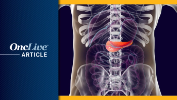
David Ilson, MD PhD: Typically, when a patient presents with esophageal or GE [gastroesophageal] junction cancer, we already have an endoscopy and biopsy to make the diagnosis. At the time of the endoscopy, we localize the tumor and identify whether the tumor is in the esophagus or GE junction or, more distally, the stomach. We generally consider esophageal and GE junction as extending up until about 2 cm into the gastric cardia. We can subtype GE junction tumors into Siewert 1, 2, and 3. Siewert 1 and 2 tumors are mainly in the esophagus/GE junction. Siewert 3 tumors are cardiac cancers with some GE junction involvement that are classified with gastric cancer.
In terms of Lauren classification, that’s really more of an issue for gastric cancer. We’re actually moving away from that now because we have more molecular-based characterization. But for distal gastric cancers, we have intestinal cancers that are more traditional adenocarcinomas regarding the association with Helicobacter pylori infection. The Lauren classification diffuse cancers are more poorly differentiated. They usually don’t form a mass. They’re usually not associated with an ulcer, and they’re more limited in the gastric wall. That being said, they’re really treated the same way, both in metastatic disease and in the surgical and adjuvant settings.
We typically use endoscopic ultrasound to identify the TNM [primary tumor/regional lymph nodes/distant metastasis] stage. We always start with a CT [computed tomography] scan to rule out distant metastatic disease, and if patients have locoregional disease with no evidence of metastatic disease, we usually do an endoscopic ultrasound to identify T and nodal staging. For gastric cancer, that’s relevant because if the tumor is T3 or node positive, we also use staging laparoscopy. About 20% of patients will have peritoneal metastasis. We don’t use that so much for esophageal and GE junction cancers because the yield is less than 5%.
Molecular subtyping is not so relevant for locally advanced disease: That’s not going to impact your treatment decisions at this juncture. But we do see that there are 4 major subtypes of esophagogastric cancer. For esophageal and GE junction cancers, 95% of these cancers are what we call genomically unstable, and that represents about 50% of gastric cancers. There’s a clear analogy between esophageal and GE junction cancers and about 50% of intestinal gastric cancers.
They share a similar molecular profile, which is that the tumors are dominated by p 53 mutations and amplification of receptor-associated tyrosine kinases, the most important of which is HER2, as well as EGFR, FGF, and MET. The other subtypes are all less common and seen more in the distal stomach setting. Those would include genomically stable tumors, which account for the large proportion of diffuse cancers. Those are relatively genomically bland. We don’t really see a lot of mutations, but we do see CDH1 somatic mutations and ROS1 mutations, which are not targetable.
Then there are 2 other very small subsets: MSI-high [microsatellite instability-high] distal gastric cancer, which usually arises from hypermethylation promoters; and, of course, one of the most important promoters, which is the silencing of the promoter that results in diminution of DNA mismatch repair proteins. You get rid of those proteins, and you get high mutation burden. Regarding MSI-high disease in a TCGA [The Cancer Genome Atlas] analysis, they reported about 20% were MSI-high. In reality, in the United States, it’s probably 10% or less. In metastatic disease, it’s probably only about 5%.
And lastly, Epstein-Barr virus—associated cancers account for about 5% to 10%. The common subtype would be genomically unstable disease, which is almost all the esophageal and GE junction tumors; 50% are gastric cancer, and then there a smattering of these other subtypes. That being said, it does not impact your decision in terms of locally advanced disease. Right now, it still has limited impact on metastatic disease. We’ll talk about the role of MSI and HER2 a little bit.
Transcript Edited for Clarity