

|Articles|December 19, 2016
- Vol. 17/No. 24
- Volume 17
- Issue 24
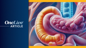
Mutational Complexity Reflects Treatment Challenges for Hepatocellular Carcinoma
Author(s)Jane de Lartigue, PhD
The growing incidence of primary liver cancer in the United States poses a great therapeutic challenge.
Advertisement
The growing incidence of primary liver cancer in the United States poses a great therapeutic challenge. Despite the plethora of new treatment options that have been tested, only a single targeted therapy is approved by the FDA for the treatment of the majority of patients who present with advanced-stage disease. Consequently, survival rates remain comparatively low.
Multiple phase III failures of promising drugs have led to the suggestion that a therapeutic plateau has been reached and that a vastly improved understanding of the molecular mechanisms of liver cancer will be necessary to drive new treatment paradigms.
Genome sequencing studies are helping to advance this goal but have highlighted key treatment challenges. In addition to the extensive heterogeneity both among and within tumors, differences in the underlying disease etiologies can significantly impact the types of potentially targetable events. Moving forward, these challenges need to be addressed in order to improve patient outcomes.
A Growing Problem
Primary liver cancer is not the most common type of cancer, but it does result in the second most common cause of cancer-related mortality worldwide, indicative of the significant treatment challenge posed by this malignancy. In recent years, the incidence of liver cancer has dramatically increased, particularly in the United States, and rates are expected to double again over the course of the next several decades.
Liver cancer typically manifests as hepatocellular carcinoma (HCC), arising from hepatocytes, which accounts for up to 85% of cases. The next most common form, intrahepatic cholangiocarcinoma (IHCC), is relatively rare in comparison; IHCC originates from the biliary tract epithelial cells in the bile duct or the liver.
The prognosis for most patients is poor, in large part due to a lack of effective surveillance techniques and limited treatment options for patients diagnosed with advanced-stage disease. Among the 30% to 40% of patients diagnosed at an early stage, surgical resection, liver transplantation, and ablative techniques offer a potentially curative option. The vast majority of patients, however, are not eligible for these treatments and advanced liver cancer is also notoriously chemo- and radioresistant, resulting in a 5-year overall survival (OS) rate of less than 10%.
The dearth of effective treatment options for these patients is not for lack of trying, but the highly complex nature of liver cancer is a major impediment to the development of new therapies. It almost always occurs against a background of underlying liver disease: liver cirrhosis is found in 80% to 90% of cases and is widely recognized as a key step in the pathogenesis of liver cancer.
Liver cancer has some of the most well-defined risk factors for any cancer. Although it is most commonly related to infection with the hepatitis B (HBV) and C viruses (HCV), other prevalent risk factors include alcohol consumption and obesity, both of which are growing concerns in developed countries in particular. The dominant risk factors vary according to geographic region and ethnic background, but all create an environment of chronic inflammation and liver damage that often leads to cirrhosis.
Major Pathways Identified
Ultimately, permanent damage to the hepatocytes prompts massive compensatory cell proliferation and regeneration, inducing genetic and epigenetic damage to the patient’s genome and fostering a highly carcinogenic environment. The process of hepatocarcinogenesis is a complex, multistep event that is still not fully understood, but several key signaling pathways have been closely linked to it.The mitogen-activated protein kinase (MAPK) and the phosphatidylinositol-3-kinase (PI3K)/Akt/ mammalian target of rapamycin (mTOR) pathways are the 2 major signaling cascades implicated in HCC development. It is also known to be a highly angiogenic tumor, with a dramatic alteration in arterial vascularity observed in many HCCs.
Thus, the MAPK and PI3K pathways, in addition to the major angiogenic pathways, have been the focus of targeted therapy development for HCC. So far, success has been limited to the multitargeted tyrosine kinase inhibitor sorafenib (Nexavar).
In the 2008 SHARP trial, sorafenib, which blocks components of both the MAPK pathway and VEGF and PDGF receptors that regulate angiogenesis, demonstrated improved OS of almost 3 months, with a manageable toxicity profile in patients with advanced-stage disease. Sorafenib has become standard of care for unresectable, nonablatable, advanced disease; however, in most cases, tumors begin to grow again after less than 6 months.
After multiple phase III failures, hopes are high that regorafenib (Stivarga) will soon join the list of approved therapies. The antiangiogenic multi-kinase inhibitor demonstrated efficacy as second-line therapy for HCC in the phase III RESORCE trial, in which median OS was 10.6 months with regorafenib plus best supportive care compared with 7.8 months for placebo plus best supportive care. This represented a 37% reduction in the risk of death (HR, 0.63; 95% CI, 0.50-0.79; P <.001).
Mutation Profile Hints at Key Drivers
Genome sequencing studies have begun to identify more of the key molecular drivers of HCC. Thousands of samples have been sequenced and the findings have implicated a number of important cellular processes and pathways in the development of primary liver cancer.
In terms of the types of mutations that are observed, similar to other solid tumors, C:G>T:A transitions are most prevalent. However, in contrast to other tumor types, T:A>C:G transitions and C:G>A:T transversions are also common, which gives HCC a unique mutation signature. Meanwhile, the frequency of mutations is similar to that of other solid tumors, averaging around 4 mutations per megabase, but there is significant variability in the exact number observed from study to study.
p53 and the Cell Cycle
Whole-genome and -exome studies have confirmed findings from earlier sequencing studies that CTNNB1 is the most frequently mutated oncogene and TP53 is the most frequently mutated tumor suppressor gene in HCC. The overall mutation frequency of CTNNB1 is 20% to 30%, but these mutations have been shown to be associated with the underlying disease etiology, with fewer CTNNB1 mutations observed in HBV-positive tumors. TP53 is mutated in 30% to 40% of HCC tumors, overall, but in contrast to CTNNB1, it is most frequently associated with HBV-positive disease.In addition to frequent mutations in the TP53 gene, genome sequencing studies have revealed that numerous other components of the p53 pathway are mutated or otherwise altered in HCC. For example, the p53-activating kinases ATM and RPS6KA3 have also been reported to be significantly mutated, although not at the frequency of the TP53 gene.
The p53 protein is a tumor suppressor whose broad function is to maintain genome stability by regulating a number of different cellular processes, including DNA repair and the cell cycle. One result of inactivating mutations in TP53, therefore, is a loss of cell cycle regulation.
Wnt/Beta-Catenin Pathway
It turns out that dysregulation of the cell cycle at the molecular level may be a characteristic of HCC, as numerous other cell cycle alterations have been detected. These include amplification of the cyclin D1 gene CCND1 and deletion of negative regulators of the cell cycle, such as CDKN2A, CDKN2B, and RB1. Overall, more than 70% of HCCs are thought to have alterations in the component genes of the p53 pathway or the cell cycle.The CTNNB1 gene encodes the beta-catenin protein, a central mediator of the beta-catenin or Wnt pathway within the cell. Vital for embryonic development, this signaling cascade has myriad roles in adult cells, reflected by its frequent association with cancer.
Telomere Stability
In addition to the CTNNB1 gene, other components of Wnt signaling are also frequently mutated in HCC. These predominantly involve the AXIN1, AXIN2, and APC genes, which negatively regulate the beta-catenin protein through posttranslational modification. It is estimated that more than 65% of HCCs show Wnt signaling-related alterations, strongly implicating this pathway in the development of HCC and presenting a potential therapeutic target.The most common mutation overall in HCC is not an activating or inactivating mutation in an oncogene or tumor suppressor, but a mutation in a non-coding part of a gene. Specifically, more than half of all HCCs display mutations in the promoter region of the telomerase reverse transcriptase (TERT) gene.
Normal cells are only able to undergo a finite number of cell divisions before they enter a senescent state, in which they no longer divide or grow. In large part, this is regulated by the length of caps on the end of the DNA, known as telomeres, which get progressively shorter with each division.
Cancer cells have acquired the capacity to maintain their telomeres. which allows them to achieve “immortality,” with an unfettered ability to grow and divide. The vast majority of cancers do this by reactivating the telomerase enzyme that is responsible for adding telomere sequences to the end of the DNA. The TERT gene encodes the catalytic core of this enzyme.
Chromatin Remodeling
Beyond TERT, a recent study looked at mutations in other noncoding regions of the HCC genome. A significant number of mutations were observed in 29 regions, implicating 6 long intergenic noncoding RNA genes, among them NEAT1 and MALAT1; 10 promoters, including those of the TFPI2, MED16, and WDR74 genes; and 9 untranslated regions (UTRs; the area just upstream of the start of a protein-coding sequence that has an important role in protein translation), including those of the BCL6 gene.Although the TP53, CTNNB1, and TERT promoter mutations represent the most commonly mutated genes in HCC, a second group of genes has been identified in genome sequencing studies. Individually, these genes are far less frequently mutated in HCC samples, but collectively they impact several key pathways.
Most numerous in this group are mutations in genes that affect chromatin remodeling. When the cell is not actively dividing, the DNA is wound tightly around histone proteins to form chromatin. Chromatin remodeling alters the way the DNA is packaged and controls the accessibility of genes for transcription, thereby regulating gene expression.
Growth Factor Signaling
Overall, alterations in genes regulating chromatin are observed in half of all HCCs. Most often, these are inactivating mutations in genes that make up the SWI/SNF chromatin remodeling complex, including ARID2, ARID1A, ARID1B, MLL, and MLL3, in addition to less frequent mutations in other components, such as SMARCA2, SMARCA4, SMARCA1, and PBRAM1. ARID2 mutations were observed more often in HCV-associated HCC, while ARID1A mutations occurred more commonly in alcohol-related cases.Another HCC group contains a large number of genes that are mutated in less than 10% of samples. This “long-tail effect,” in which the vast majority of significantly mutated genes occurs at low frequencies, is common across tumor types, particularly in those with substantial mutational heterogeneity. It represents one of the most significant issues limiting the development of targeted therapy.
Interestingly, among HCCs, this group of mutations appears to be made up of many genes involved in growth factor signaling. This could help to explain the apparent promise of targeting growth factor signaling pathways in HCC, but the difficulties experienced in achieving success in the clinic thus far.
Alterations in drug target kinases are rare and mutations in growth factor signaling are mostly represented by those in the FGFR2, FGFR3, KIT, and JAK1 genes, as well as amplification of the MET gene. Activating mutations in the RAS, PIK3CA, and EGFR genes are rare and, when mutations are observed, they typically are not the same types of mutations commonly observed in other tumor types.
EGFR gene amplification and protein overexpression is also rare in HCC, yet the EGFR, RAS, and PI3K pathways have all been demonstrated to play a role in the development of HCC. Exactly how they are involved, however, remains unclear, although there are suggestions they may be activated in other ways.
HBV Integration
For example, mutations were observed in the ERRFI1 gene, which encodes the ERBB receptor feedback inhibitor 1, a protein that inhibits the kinase activity of EGFR and HER2. Thus, this mutation may remove that inhibitory activity and drive activation of these pathways. In addition, mutations in the TSC1 and TSC2 genes that could activate the mTOR protein have also been reported.More than 80% of HCCs are associated with HBV or HCV infection, with HBV being most common worldwide. Since these are DNA viruses, one of the ways in which they drive HCC development is by integrating their viral DNA into the host genome. In a manner similar to other types of genetic damage, this can alter the expression and function of host genes and induce genomic instability.
Several genome sequencing studies have begun to examine the consequences of HBV integration. Among the hundreds of integration sites that have been identified, there is a significant overrepresentation of coding or promoter regions, suggesting that HBV integration confers a growth advantage by regulating host gene expression.
Cell Cycle, Adhesion, and Oxidative Stress
The most common integration sites involve the TERT, MLL4, and CCNE1 genes, strongly implicating telomere stability and the cell cycle in the development of HCC. These integration events were shown to significantly increase the transcription and expression of these genes. In addition, the number of HBV integration sites was significantly associated with more aggressive disease and poorer prognosis.Other categories of mutations that have been identified include dysregulation of the cell cycle at the G1/S checkpoint through inactivation of the RB and CDKN2A genes, and aberrations in genes that regulate adhesion molecules involving the epithelial cells (EpCAM).
Wide-Ranging Implications of Heterogeneity
Additonally, there is oxidative stress resulting from an imbalance between the generation of reactive oxygen species and the body’s ability to neutralize them or repair the resulting damage. When this occurs, cells orchestrate an adaptive response that functions to counter the effects and restore balance.By unraveling the genomic alterations underlying HCC, a picture of significant heterogeneity has emerged, with only a few common driver mutations identified in the majority of patients and a plethora of genes that are infrequently mutated.
Studies have also shown that HCC develops through clonal evolution, resulting in significant heterogeneity even within the same tumor. Although the majority of abnormalities are found across multiple regions of a single tumor, representing a common genetic basis for tumor development, each tumor region also acquires independent mutations.
Heterogeneity has important implications for the development of new drugs and is likely one of the principal reasons that genomic findings have not, as of yet, translated into any significant therapeutic progress. It also impacts the development of biomarkers if these molecular alterations are not identified uniformly across or within tumors as this may lead to differential responses to therapy.
KEY RESEARCH
Fujimoto A, Furuta M, Totoki Y, et al. Whole-genome mutational landscape and characterization of noncoding and structural mutations in liver cancer. Nat Genet. 2016;48(5):500-509. doi:10.1038/ng.3547.
Fujimoto A, Totoki Y, Abe T, et al. Whole-genome sequencing of liver cancers identifies etiological influences on mutation patterns and recurrent mutations in chromatin regulators. Nat Genet. 2012;44(7):760-764. doi:10.1038/ng.2291.
Llovet JM, Villanueva A, Lachenmayer A, Finn RS. Advances in targeted therapies for hepatocellular carcinoma in the genomic era. Nat Rev Clin Oncol. 2015;12(7):408-424. doi:10.1038/nrclinonc.2015.103.
Cancer Research Institute. Timeline of progress. http://www.cancerresearch.org/our-strategy-impact/timeline-of-progress/ timeline-detail. Accessed November 28, 2016.
Nakagawa H, Shibata T. Comprehensive genome sequencing of the liver cancer genome. Cancer Lett. 2013;340(2):234-240. doi:10.1016/j.canlet.2012.10.035.
Ozen C, Yildiz G, Dagcan AT, et al. Genetics and epigenetics of liver cancer. N Biotechnol. 2013;30(4):381-384. doi:10.1016/j.nbt.2013.01.007.
Shibata T, Aburatani H. Exploration of liver cancer genomes. Nat Rev Gastroenterol Hepatol. 2014;11(6):340-349. doi:10.1038/nrgastro.2014.6.
Totoki Y, Tatsuno K, Covington KR, et al. Trans-ancestry mutational landscape of hepatocellular carcinoma genomes. Nat Genet. 2014;46(12):1267-1273. doi:10.1038/ng.3126.
Articles in this issue
about 9 years ago
A Time for Hopeabout 9 years ago
Expert Discusses Recent Progress in NET Managementabout 9 years ago
Targeted Therapy Makes Inroads in Gastroesophageal Cancerabout 9 years ago
Tepotinib Takes Aim at Novel MET Mutation in Lung Cancer Trialabout 9 years ago
Patient-Friendly Policies Extend to Decision MakingAdvertisement
Related Content
Advertisement


Latest CME


Advertisement
Advertisement
Trending on OncLive
1
Single-Center, Retrospective Data Show Low Rate of Lifileucel Infusion Following Referral in Advanced Melanoma
2
Outcomes With Bridging Therapy Correlate With Cilta-Cel Efficacy, Safety in Multiple Myeloma
3
Real-World Data Support Clinical Benefit With Lifileucel in Previously Treated Advanced Melanoma
4
Long-Term Cilta-Cel Data Show Low Rates of PFS Events in Standard-Risk R/R Myeloma
5