New data on multiple targeted agents and evolving diagnostic frameworks highlight progress in classifying and treating EGFR PACC–mutated NSCLC.
Computed tomography-based image guidance during radiation therapy could be a method of identifying patients who require a complete workup for COVID-19.
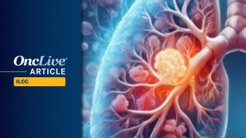
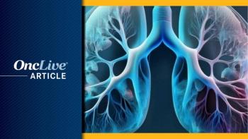
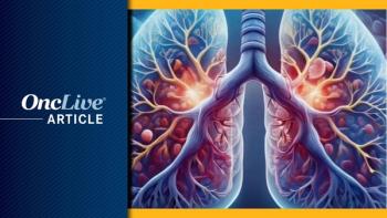
Computed tomography (CT)-based image guidance during radiation therapy could be a method of identifying patients who require a complete workup for COVID-19, according to findings from an analysis of patients with lung cancer published in the Journal of Thoracic Oncology.1,2
“These findings are extremely important,” lead study author Graham Warren, MD, PhD, a professor at the Medical University of South Carolina, said in a press release. “If a doctor notices something on the patient’s scan, they can have them tested for COVID-19 or ask them how they are doing. This gives us the opportunity to identify patients who may have a higher risk of having COVID-19, whether they are symptomatic or not.”
Screening for COVID-19 exposure, particularly for patients with cancer on active treatment, has become a priority during the pandemic. Following 2 case reports of imaging findings during routine CT-based image-guided radiation therapy scans, investigators set out to determine whether there is a correlation between the onset of new pulmonary infiltrates found during radiation therapy in patients diagnosed with COVID-19.
To that end, investigators compiled data of deidentified case reports of patients who developed biochemically confirmed COVID-19 during radiation therapy through an online portal. The data included information on the patient’s sex, age, cancer diagnosis and treatment, and COVID-19 diagnosis and outcome.
“Lung cancer patients receiving radiation are unique because they are already getting routine lung scans,” Warren said. “We can use the radiation setup already in place to identify risk of COVID-19, and we can accomplish this without significant changes to our standard radiotherapy procedures.”
During the series, coplanar CT-based imaging was performed on each patient to identify the presence or absence of ground-glass opacities or infiltrates.
“We want to try to identify a cancer patient’s risk of having COVID-19 very quickly because these patients have a higher risk of complications and mortality,” Warren said. “By using scans to discover potential positive COVID-19 cases early on, we can help to decrease the risk of exposure to other patients as well as facilitate earlier diagnosis and treatment for patients who have COVID.”
Seven cases were submitted from Turkey, Spain, Belgium, Egypt, and the United States. These cases were supplemented by results and imaging from 2 patients reported by Suppli et al and McGinnis et al.
The median patient age was 63.3 years.
The 9 patients included 4 females and 1 patient with metastatic disease. All 9 patients had lung cancer, including 4 cases of squamous cell carcinoma and 5 cases of adenocarcinoma.
All patients had confirmed COVID-19 diagnoses using polymerase chain reaction–based tests or nasopharyngeal swabs.
Regarding cancer treatment characteristics, 7 patients received chemotherapy and 1 patient was enrolled in a clinical trial evaluating immunotherapy plus stereotactic ablative radiation therapy.
The results showed that abnormalities indicative of ground-glass opacities or infiltrates were identified in 8 of the 9 patients analyzed. The ninth patient did not have any evident opacities or infiltrates on submitted images.
Three patients completed the planned course of radiation therapy; the remaining patients interrupted or discontinued treatment.
Six patients experienced symptoms, with fever presenting as the most common symptom in all 6 patients.
Seven patients were hospitalized, including 1 patient who was hospitalized without symptoms because of standard local protocol.
As of the date of data submission, 2 patients had died.
“With current COVID-19 variants and symptoms, these imaging findings could be extremely useful to help to minimize risk and infection,” Warren said. “These findings directly align with strategies to identify optimal cancer treatment strategies coupled with harm reduction strategies for COVID-19. These simple findings can reduce risk and exposure and can immediately improve the lives of cancer patients during cancer treatment, Warren concluded.”