|Videos|January 24, 2023
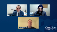
Workup and Molecular Testing for a Patient with NSCLC
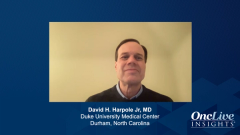
David H. Harpole Jr, MD, a thoracic surgeon, details his workup approach and discussed molecular testing for patients who present with NSCLC.
Advertisement
Episodes in this series
Transcript:
David Spigel, MD: David, I’m curious. When I started almost 20 years ago out of training, I remember the surgeons I worked with and they were great. Their advice was, if it’s a solitary lesion, just take it out, don’t even bother biopsying it. Of course, back then we were just learning how to do PET [positron emission tomography] scans. [Discuss] your thoughts on how a patient who comes to you at Duke [University Medical Center] approaches with a solitary lesion. What are you doing? Are you doing a PET scan? If I’m not mistaken, I think you do your own scopes, but could you expand on your approach to the workup of a solitary lesion?
David H. Harpole Jr, MD: It has certainly evolved, and frankly, most rapidly over the last 2 to 4 years. As this wave is moving across the country and things are changing, I will tell you, we were a universal mediastinoscopy center for everyone. I used to do literally 300 or 400 a year, and now I do like 15…. I think the EVIS EXERA III platform that we use, and interventional pulmonary uses, has supplanted that. I will say that with the robotic platforms now, I think that has been our primary [procedure] as well. The reason being is that historically, [with] a 2-cm lesion like this that’s suspicious, we would’ve just taken it out without a diagnosis. We can go more into about the size and what we find, but certainly lesions greater than 3 cm where there are some other options now, we have been getting not only cytology but histology and molecular information on them, and we’re going to obviously discuss that more.
This [case] is pretty much a straightforward T1 lesion that I would say probably half of the thoracic surgeons now would take out without a diagnosis. We tend to get a diagnosis now more than we used to, and I think it has killed 2 birds with 1 stone because with EVIS EXERA III and our navigational [bronchoscopy], we can get a tissue diagnosis of the lesion as well as the nodal stations at the same time. This is truly a team [effort], and I think our interventional pulmonary team has become even more important to that.
Transcript edited for clarity.
Advertisement
Related to this article

Panelists discuss how nodal burden, NGS turnaround time, and regional resource heterogeneity shape treatment sequencing in EGFR-mutated NSCLC in Latin America.

In a recent OncLive Peer Exchange, expert investigators discussed the utility of ctDNA and MRD testing in solid tumors.

The European Union approved trastuzumab deruxtecan for previously treated, HER2-postive metastatic solid tumors.
SB27, a proposed pembrolizumab biosimilar, met primary end points in both a phase 1 pharmacokinetic and a phase 3 efficacy study.
OS was not improved with sigvotatug vedotin vs docetaxel in patients with pretreated, locally advanced, unresectable, or metastatic nonsquamous NSCLC.
Advertisement
Advertisement
Trending on OncLive
1
Managing Long-Term Toxicities and Emerging Therapies in ALK-Positive NSCLC
2
Five Under 5: Top Oncology Videos for the Week of 6/28
3
The OncFive: Top Oncology Articles for the Week of 6/28
4
Divesiran Reduces Phlebotomy Burden and Improves Iron Stores in Polycythemia Vera
5