|Videos|March 1, 2021
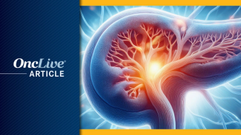
Cholangiocarcinoma: Appropriate Diagnostic Work-Up
Types of assessments that are used to help appropriately work up a patient suspected of having a bile duct cancer.
Advertisement
Episodes in this series


John L. Marshall, MD: Let’s look at the work-up and overview of this disease. I’m always impressed with our colleagues in surgery and GI [gastrointestinal] who are charged with diagnosing, particularly extrahepatic and Klatskin or hilar, tumors. Milind, do you want to walk us through that original presentation diagnosis, the role of gene testing, etc?
Milind Javle, MD: Klatskin tumors—also known as perihilar tumor, or hilar cholangiocarcinoma, extrahepatic cholangiocarcinoma—are complicated. These patients present with jaundice. There’s a subtle mass or none at all. There’s a malignant-looking stricture, and the ERCP [endoscopic retrograde cholangiopancreatography] comes back with cytology as nondiagnostic. It’s only 20% to 25% of the time that cytology is useful. If you have an associated lymph node, then you can get an endoscopic ultrasound and a biopsy. However, there’s a substantial proportion of patients—perhaps one-third—for whom the diagnosis is made on clinical grounds, where you have a mass, a stricture, and an elevated
CA [cancer antigen] 19-9. The consensus is that this is perihilar cholangiocarcinoma. These patients unfortunately go through investigation after investigation to come to the diagnosis.
The diagnosis of intrahepatic cholangiocarcinoma is relatively straightforward. You have a mass, or an infiltrate. If it’s amenable to needle biopsy, it is done, and then a diagnosis can be made. The diagnosis is one of exclusion, as we’ll discuss later. Immunohistochemical profile helps us narrow it down. We don’t need a lot of unnecessary diagnostic steps with a good immunohistochemical picture there.
John L. Marshall, MD: I struggle with this, particularly with the difficult-to-diagnose that you referred to. I felt crazy a year and a half or more ago when we were in tumor board, and they kept trying to do brushings that were stented; surgeons don’t want to act without a diagnosis. We don’t want to give chemotherapy or radiation without a diagnosis. I raised my hand and said, “Could we do a circulating test? If we found a cancer mutation, maybe that’s enough.” Where do you all think that is? I know there was an abstract; our friend Pashtoon Kasi published an abstract at ASCO GI [American Society of Clinical Oncology Gastroinstestinal Cancers Symposium] about this. Do you all think that’s appropriate? Is that enough? Is that just a lead? How do you incorporate that kind of testing?
Sameek Roychowdhury, MD, PhD: That’s a good question. Can we use a cell-free DNA test at initial diagnosis to say it’s cancer? That’s an interesting proposition. The challenge could be that not all intrahepatic or hilar cholangiocarcinomas are going to have a mutation on such a cell-free DNA test. The cell-free DNA tests that we have today are not as big in terms of the number of genes. If you don’t have a mutation represented on one of those gene panels from one of our current liquid biopsies, you may miss a diagnosis. Only a fraction of the patients with cholangiocarcinoma will have such a mutation; we can talk about the breakdown of those genes that we do expect.
The other thing is that not all cell-free DNA tests are going to adequately represent someone’s tumor. A characteristic that affects a liquid biopsy is the tumor site, so what organism infiltrated the tumor burdens. You can have a small tumor that is obstructing a duct, or in the distal duct, but does not have a large tumor burden, so you may not see a lot of cell-free material. The mutations may not catch something. Those 3 things can end up leading you to a false-negative. If positive, it is helpful, but if it’s negative, it’s not necessarily going to be helpful for early diagnosis.
I completely agree with Milind, but there could be a diagnostic escapade of things that happened to make the diagnosis. I am particularly pushy about it because we need the tissue at some point. The diagnosis can be made clinically. Sometimes you need to get someone started on treatment because they’re having a lot of trouble, but the tissue is really critical because it can help guide us for future therapies that are quite beneficial. I think we’ll talk about them today. Without that tissue, it’s hard to make such testing possible. While cell-free DNA is available, there are some caveats. I’m one of the biggest proponents of cell-free DNA as a tool and research tool at the moment, but a few things are going to get missed for various reasons: tumor burden, organ site effects, and whether you’ve even got the right probes for your test to capture the alteration. I worry about missing things that could really help people. I would never want to miss microsatellite instability. Even a 5% miss is a big miss, in my view, for microsatellites.
John L. Marshall, MD: I keep coming back to the tissue-informed cell-free DNA testing vs just casting a net. A negative doesn’t really help you, but if you had a positive, it would at least give us enough to say cancer or not. Katie, what would be a standard on the West Coast for this approach?
R. Kate Kelley, MD: We don’t use it routinely as part of diagnosis. I’ll admit, in some of those difficult cases where on the nth endoscopic attempt at a SpyBite intraductal biopsy or US biopsies, we keep getting atypical or suspicious results but nothing confirmatory. I have sent it before and felt reassured that when I see a KRAS mutation, if there is a tumor with all the caveats that Sameek shared, it doesn’t confirm the primary sites. I may still have to infer that from the anatomy. It is not validated as a diagnostic test. In tumor board, there are cases where we try extremely hard, but I have seen us wait too long to start therapy, being uncomfortable with strong suspicions. Sometimes we have to make a clinical decision.
Transcript Edited for Clarity
Advertisement
Related to this article

The FDA approved Orca-T, issued a CRL to a NET imaging agent, and more.

Adagrasib in combination with cetuximab did not significantly improve OS or PFS in KRAS G12C–mutated metastatic colorectal cancer.

The FDA gave a CRL to the NDA seeking the approval of Orviglance for use in the detection of liver metastases in patients with severe kidney impairment.
Updated analyses from a phase 2a study suggest durable survival with namodenosen in a subset of heavily pretreated patients with advanced PDAC.
Need a refresh on the current regulatory landscape for GI oncology? We've got you covered. Here's everything that moved the needle across GI malignancies this month.

The FDA has issued a CRL to LNTH-2501 for the localization of SSTR-positive NETs.

The European Union approved trastuzumab deruxtecan for previously treated, HER2-postive metastatic solid tumors.
Advertisement
Advertisement
Trending on OncLive
1
Epcoritamab Plus Lenalidomide/Rituximab Win EU Approval for R/R Follicular Lymphoma
2
TP53 Status and Time Toxicity Shape Sequencing Decisions in EGFR-Mutated NSCLC
3
Divarasib Outperforms First-Generation KRAS G12C Inhibitors in Phase 3 NSCLC Trial
4
Savolitinib Gains NMPA Approval for MET-Amplified Gastric and GEJ Adenocarcinoma
5