|Videos|July 16, 2021
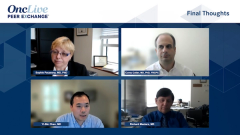
Diagnosing & Staging of GVHD
Sophie Paczesny, MD, PhD, leads a discussion on diagnostic tests used in the setting of graft-vs-host disease and how patients are risk stratified. Whether organ biopsies are used routinely is also discussed.
Advertisement
Episodes in this series
Sophie Paczesny, MD, PhD: Is organ-specific biopsy always recommended? Is that still the gold standard? When I looked at retrospective studies, only 70% were really correlated to GVHD [graft-vs-host disease]. What are your thoughts on this?
Corey Cutler, MD, PhD, FRCPC: We do not mandate biopsies. Certainly, they’re useful to exclude competing pathologies, for example CNB colitis. We biopsy the liver the least. The findings in skin biopsies are generally very non-specific; even a skin biopsy in the presence of an offending drug agent that might be causing an allergic type of rash might not be able to be easily differentiated on a biopsy. I would say other than the GI [gastrointestinal] tract, which we basically biopsy on everybody, we do very few other biopsies.
Yi-Bin Chen, MD: I’d agree with that. We are speaking mainly right now of acute GVHD. Liver being the most invasive and thus the riskiest, we definitely biopsy the least. At the same time, if it’s isolated liver disease, which is the rarest of the bunch here, and depending on how high the LT abnormalities are, you often feel the need to do that; otherwise it’s tough to figure it out if it’s isolated liver GVHD. Otherwise, I agree; we rarely biopsy the skin unless a clinical trial mandate it. However, gut biopsies we frequently do.
Sophie Paczesny, MD, PhD: I notice you do not do repeat biopsies, right? I have seen that in Europe; they have protocols. Sometimes you do repeat biopsies if the first one was not complementary or sometimes to see the response. I do not think this is usual in centers in the US, correct?
Corey Cutler, MD, PhD, FRCPC: We will do repeat GI biopsies before initiating subsequent lines of therapy just to ensure that there isn’t intercurrent infectious pathology as well. That’s the one time I do repeat biopsies.
Yi-Bin Chen, MD: I agree. Some studies, people feel that you can often look at a GI biopsy and see if there’s healing underway and regenerating mucosa; I’m not so sure how reliable that is. I think there’s a lot of sampling bias involved in a random GI biopsy. For the most part, it’s done to exclude something on top of it. We don’t uncommonly do it, but that’s not saying we commonly do it.
Sophie Paczesny, MD, PhD: It’s more for differential diagnosis, too, and sometimes for response. Another question is how early do you do the biopsy? As soon as there are signs? I know that, for instance, Fred Hutch [Cancer Center] has an approach where they scope almost everyone with a little nausea, vomiting, and we have a higher grade of GVHD. The usual approach is to wait a little bit, correct?
Corey Cutler, MD, PhD, FRCPC: Yes. For us, do not do a lot of upper GI endoscopy for nausea, vomiting, or bloating. That needs to be a persistent sign or symptom before we will approach that as a diagnostic entity. I think there’s a lot of gut apoptosis in the context of conditioning injury; one can be a little bit confused if one does early biopsies when patients haven’t quite recovered from their conditioning-related toxicities.
Richard Maziarz, MD: I do think it’s important to note that you may not always get immediate service from GI specialists if your patient is on a study or if you so choose to get a biopsy, particularly in the case of GI tract GVHD. It may be 24 to 48 hours before they can find the time to build in their schedule to come do this; we don’t wait to treat. The important take-home is, you may look for biopsy for information for confirmation, but if you clinically suspect it the worst thing you can do is hold off treatment. By then, you’ll have 2- or 3- or 4-log further expansion of the T-cell clones that are causing the pathology.
Sophie Paczesny, MD, PhD: We talked about differential diagnosis only using imaging. There is new technology where you can check the skin changes or also some imaging actually for the GI tract. I’m not sure if you’re using this and if you have seen advantage of using this.
Yi-Bin Chen, MD: We are not using imaging specifically for the diagnosis or monitoring of progress of GVHD. At the same time, it is common for someone who is hospitalized for GI GVHD to have imaging of the abdomen at some point due to the complaints that they have; you’re trying to rule out other things and the abdominal exam is such a black box. We all know there are specific CT findings that are helpful in some ways; either the scattered diffuse inflammation or in certain cases the isolated persistent terminal ileal thickening that remains very difficult to treat. In the rare patient, we’re able to get PET imaging; that’s usually in a patient that has a disease that needs to be restaged, or the PET CT such as lymphoma. PET imaging can be really helpful if we use it correctly for this disease as certain studies have shown. I’m just not sure we’re able to apply it at the moment with issues of access and reimbursement and so forth. I think there will be more non-invasive ways in the future where we’re able to monitor the progress of this disease.
Richard Maziarz, MD: What I think is difficult in the case of making a diagnosis as we talk about GI tract, and even if we get into lung where we talk about pulmonary chronic GVHD, is when someone has a preexisting infection. We sometimes will go back to prove we don’t have a super infection. However, there is also the circumstance where we may have CMV colitis or we may have Clostridium difficile colitis that leads to a syndrome of GI tract distress that looks like GVHD, and then it may be improving and then they worsen. Are they worsening because they are relapsing their infectious disease process, or have we now created an inflammatory state that leads to the subsequent secondary activation of graft-vs-host? I think that the biologies are intermixed; on a clinical level, you have to maintain that high level of suspicion and inquiry, not to assume that a single diagnosis may explain all.
Sophie Paczesny, MD, PhD: What about chronic GVHD, like Corey and Yi-Bin said at the beginning?
It is far more complex. My understanding is that we try to avoid biopsy, the skin biopsies, for chronic GVHD in most cases. In my center where I was before and in my new center, we try to completely avoid lung biopsies, for instance. Comments on this?
Corey Cutler, MD, PhD, FRCPC: Again, it’s largely a clinical diagnosis. The pathologic findings can be very variable in chronic graft-vs-host disease. Many of the tissues that you’re interested in are not terribly accessible, other than the skin which has such classic clinical presentation, that often biopsy is really not necessary. At that point in the patient’s course, there is really not a lot that’s in the differential diagnosis. For that reason, we do very few biopsies unless it is mandated by the clinical trial.
There are certain trials that are looking at correlative biomarkers in tissue, such as in the mouth or in saliva, where you can get some samples. However, we try not to do too many biopsies in this scenario as well.
Sophie Paczesny, MD, PhD: Now for the diagnosis and for staging. Acute GVHD is relatively easy, correct? I suppose if you have old clinics, again that big center, in a community center, it’s a little bit more difficult to do this extensive chronic GVHD evaluation. Does anyone have any insight on this? I think it takes at least 30 minutes of somebody with expertise, right, like might not be possible at a community center.
Corey Cutler, MD, PhD, FRCPC: I’m not sure about 30 minutes. I think if you have those 3 pages printed out in advance, not everyone is going to have every organ involved. I think you can probably do a thorough enough chronic GVHD exam in under 10 minutes. Obviously, we do not examine eyes terribly well as clinicians, but there is no examination component to the eye. A skilled clinician can do a proper chronic GVHD assessment in about 10 minutes or so I would say, if you are familiar with what needs to be measured.
Yi-Bin Chen, MD: It does depend on how many organs are involved. I would say that the NIH [National Institute of Health] criteria, while it’s the best we have and we use it for clinical trial endpoints and such metrics, it is a bit difficult to use in the workflow of a busy clinic. I think all of us would agree that we are not using the NIH criteria on a routine basis off trials to make decisions about patients while we are sitting with the patient themself. That is because it takes a long time to do. It takes time to do that entire assessment properly, and you are trying to move with the patient and make decisions and so forth. There is a disconnect there in the tool we use to judge clinical trials and the tools we use to make direct decisions about patients daily. I will agree with that, absolutely.
Sophie Paczesny, MD, PhD: What are some of the shortcomings? If you refocus on acute GVHD, so beyond ..... for restratification, still we have come with good 2 biomarkers. Plasma biomarker are interesting, at the time where predictive biomarkers or at the time of onset of GVHD to say that the patient at high risk or low risk. This has led to a clinical trial that was quite interesting…and you have all participated, too. When you look at the standard risk patient, they were randomized between sirolimus and the standard of care that’s corticosteroids, the outcomes are similar between the 2 groups. That was an interesting study; saying that the clinical grade plus the biomarkers can help change outcomes or treatment. This is the only trial that is out there saying that predictive biomarkers could be interesting. It has been tried on the higher-risk patient. The clinical trial is still underway. Nothing can be randomized because people think it is not ethical to have a randomized trial for high-risk patients. The program is probably [good] but we do not have the right treatment yet. So far there is no real difference in the high-risk group. When you can also try a preemptive trial, where you look at the risk biomarkers before clinical signs and see if you can diffuse basically an actual GVHD ….. There is no clinical trial yet and algorithms are probably not as precise as predictive biomarkers; I think that could be something that needs to be done in the future. You must balance the risk and the benefit. You must target the high-risk group for this. I believe that is the future; I’m really interested in doing this ….. I know that other people have tried to do this. Sometimes the problem is: which treatment do you use for this? We have done this with alpha antitrypsin and that has not been successful. But probably they blame ..... alpha antitrypsin, it might be also because of the biomarker was not specific and sensitive enough. We have to find also really good ..... to target, we need only for high-risk population.
For acute GVHD, I don’t know if you have questions about this, that’s where we stand so far. I think the best call right now is predictive biomarkers, and we can also monitor the response. That has been quite good; when you look at the biomarker and if there is a response, it should decrease.
Yi-Bin Chen, MD: The acute GVHD biomarkers that Sophie mentioned, ST2 and REG3-alpha, are available commercially. We do not send them right now outside of a clinical trial. I’m just wondering, does anybody send them right now for prognostic purposes or to make decisions?
Corey Cutler, MD, PhD, FRCPC: We do not.
Sophie Paczesny, MD, PhD: In pediatric, some pediatricians are sending it when they have a really high suspicion. The problem with the algorithm is: you don’t know really what is in there, right? You have an algorithm and you need to be sure to use the right one. If it’s at onset of GVHD, it’s a different algorithm from the one early on. That might be a little bit confusing for the clinician, right?
Yi-Bin Chen, MD: Yeah.
Sophie Paczesny, MD, PhD: I think that has been a limiting factor for the biomarkers.
Richard Maziarz, MD: There is another circumstance which is we focus on that first diagnosis of whether it’s acute or the first diagnosis of chronic. I had a patient I saw last week who creates a diagnostic dilemma where she has had scleroderma chronic GVHD for 10 years. We transplanted her 15 years ago and she has now been off all immune suppressive medicines for over half a decade. In the last 3 to 6 months, she is now having pulmonary findings and also may have more sclerosis now extending up the lateral aspects of her chest. We do not really think about ongoing sclerodermatous progression; I think that would be another valuable tool. We spend our time trying to figure out who’s going to get it, but what do we do for the people who have it?
Yi-Bin Chen, MD: With those types of patients, it’s always a question of what’s reversible and what’s not reversible. It would be helpful to have some sort of biological biomarker to help us understand that; to help you figure out how aggressive to be for these, especially if you are out 10 or 15 years. That is really tough.
Transcript edited for clarity.
Advertisement
Related to this article

Teclistamab plus talquetamab met the primary PFS end point and improved overall survival in relapsed/refractory multiple myeloma in MonumenTAL-6.

The FDA has granted priority review to a supplemental application for talazoparib plus enzalutamide in HRR-altered mCSPC.
Camizestrant plus a CDK4/6 inhibitor was approved in the European Union for ER-positive, HER2-negative advanced breast cancer with an emergent ESR1 mutation.
The FDA is reviewing data for daraxonrasib in previously treated pancreatic cancer under the Commissioner’s National Priority Voucher program.
Johannes Schetelig, MD, MSc, discusses phase 3 data for post-transplant cyclophosphamide vs ATLG as GVHD prophylaxis in hematologic malignancies.
Matthew Galsky, MD, discusses the FDA approval of perioperative pembrolizumab plus enfortumab vedotin in MIBC.

The FDA has approved zidesamtinib in patients with locally advanced/metastatic ROS1+ NSCLC who received a prior ROS1 kinase inhibitor.
A 3-year, $225K award will focus on improving quality of life for patients living with long-term effects of cancer treatment.
Advertisement
Advertisement
Trending on OncLive
1
FDA Accepts NDA for Daraxonrasib in Previously Treated Metastatic Pancreatic Cancer
2
FDA Approves Zidesamtinib in Pretreated ROS1+ NSCLC
3
Dr Galsky on Systemic Therapy Advances in Muscle-Invasive Bladder Cancer
4
Dr Patel on the Unmet Need in EGFR+ Transformed Small Cell Lung Cancer
5